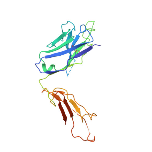
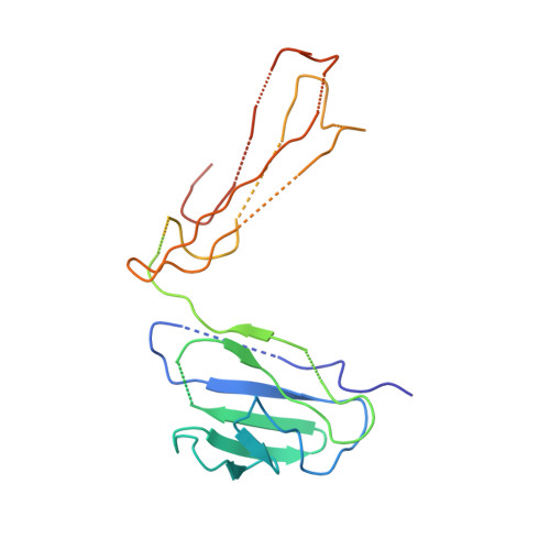
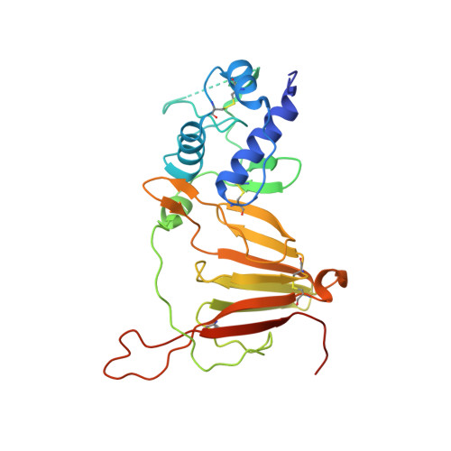
8DDK

CCHFV GP38 Hoti/Kosovo bound with CC5_17


Experimental Data Snapshot

  • Method: X-RAY DIFFRACTION
  • Resolution: 3.86 Å
  • R-Value Free: 0.335 
  • R-Value Work: 0.304 
  • R-Value Observed: 0.305 

Starting Model: experimental
View more details

wwPDB Validation   3D Report Full Report


This is version 1.1 of the entry. See complete history


Literature

Structural characterization of protective non-neutralizing antibodies targeting Crimean-Congo hemorrhagic fever virus.

Durie, I.A.Tehrani, Z.R.Karaaslan, E.Sorvillo, T.E.McGuire, J.Golden, J.W.Welch, S.R.Kainulainen, M.H.Harmon, J.R.Mousa, J.J.Gonzalez, D.Enos, S.Koksal, I.Yilmaz, G.Karakoc, H.N.Hamidi, S.Albay, C.Spengler, J.R.Spiropoulou, C.F.Garrison, A.R.Sajadi, M.M.Bergeron, E.Pegan, S.D.

(2022) Nat Commun 13: 7298-7298

  • DOI: https://doi.org/10.1038/s41467-022-34923-0
  • Primary Citation of Related Structures:  
    8DC5, 8DCY, 8DDK

  • PubMed Abstract: 

    Crimean-Congo Hemorrhagic Fever Virus (CCHFV) causes a life-threatening disease with up to a 40% mortality rate. With no approved medical countermeasures, CCHFV is considered a public health priority agent. The non-neutralizing mouse monoclonal antibody (mAb) 13G8 targets CCHFV glycoprotein GP38 and protects mice from lethal CCHFV challenge when administered prophylactically or therapeutically. Here, we reveal the structures of GP38 bound with a human chimeric 13G8 mAb and a newly isolated CC5-17 mAb from a human survivor. These mAbs bind overlapping epitopes with a shifted angle. The broad-spectrum potential of c13G8 and CC5-17 and the practicality of using them against Aigai virus, a closely related nairovirus were examined. Binding studies demonstrate that the presence of non-conserved amino acids in Aigai virus corresponding region prevent CCHFV mAbs from binding Aigai virus GP38. This information, coupled with in vivo efficacy, paves the way for future mAb therapeutics effective against a wide swath of CCHFV strains.


  • Organizational Affiliation

    Department of Pharmaceutical and Biomedical Sciences, College of Pharmacy, University of Georgia, Athens, GA, USA.


Macromolecules
Find similar proteins by:  (by identity cutoff)  |  3D Structure
Entity ID: 1
MoleculeChains Sequence LengthOrganismDetailsImage
Heavy Chain CC5-17243Homo sapiensMutation(s): 0 
Entity Groups  
Sequence Clusters30% Identity50% Identity70% Identity90% Identity95% Identity100% Identity
Sequence Annotations
Expand
  • Reference Sequence
Find similar proteins by:  (by identity cutoff)  |  3D Structure
Entity ID: 2
MoleculeChains Sequence LengthOrganismDetailsImage
Light Chain CC5_17212Homo sapiensMutation(s): 0 
Entity Groups  
Sequence Clusters30% Identity50% Identity70% Identity90% Identity95% Identity100% Identity
Sequence Annotations
Expand
  • Reference Sequence
Find similar proteins by:  (by identity cutoff)  |  3D Structure
Entity ID: 3
MoleculeChains Sequence LengthOrganismDetailsImage
Envelopment polyprotein274Orthonairovirus haemorrhagiaeMutation(s): 0 
UniProt
Find proteins for B2BSL7 (Orthonairovirus haemorrhagiae)
Explore B2BSL7 
Go to UniProtKB:  B2BSL7
Entity Groups  
Sequence Clusters30% Identity50% Identity70% Identity90% Identity95% Identity100% Identity
UniProt GroupB2BSL7
Glycosylation
Glycosylation Sites: 2
Sequence Annotations
Expand
  • Reference Sequence
Oligosaccharides

Help

Entity ID: 4
MoleculeChains Length2D Diagram Glycosylation3D Interactions
alpha-D-mannopyranose-(1-2)-alpha-D-mannopyranose-(1-3)-beta-D-mannopyranose-(1-4)-[alpha-D-mannopyranose-(1-3)]2-acetamido-2-deoxy-beta-D-glucopyranose-(1-4)-2-acetamido-2-deoxy-beta-D-glucopyranose
D
6N-Glycosylation
Entity ID: 5
MoleculeChains Length2D Diagram Glycosylation3D Interactions
alpha-D-mannopyranose-(1-3)-beta-D-mannopyranose-(1-4)-2-acetamido-2-deoxy-beta-D-glucopyranose-(1-4)-2-acetamido-2-deoxy-beta-D-glucopyranose
E
4N-Glycosylation
Glycosylation Resources
GlyTouCan:  G81315DD
GlyCosmos:  G81315DD
GlyGen:  G81315DD
Experimental Data & Validation

Experimental Data

  • Method: X-RAY DIFFRACTION
  • Resolution: 3.86 Å
  • R-Value Free: 0.335 
  • R-Value Work: 0.304 
  • R-Value Observed: 0.305 
  • Space Group: P 43 21 2
Unit Cell:
Length ( Å )Angle ( ˚ )
a = 175.676α = 90
b = 175.676β = 90
c = 92.158γ = 90
Software Package:
Software NamePurpose
PHENIXrefinement
HKL-2000data reduction
PDB_EXTRACTdata extraction
HKL-2000data scaling
PHENIXphasing

Structure Validation

View Full Validation Report


Entry History & Funding Information

Deposition Data


Funding OrganizationLocationGrant Number
National Institutes of Health/National Institute Of Allergy and Infectious Diseases (NIH/NIAID)United StatesR01AI109008
National Institutes of Health/National Institute Of Allergy and Infectious Diseases (NIH/NIAID)United StatesR01AI151006
Defense Threat Reduction Agency (DTRA)United StatesHDTRA12110005

Revision History  (Full details and data files)

  • Version 1.0: 2022-12-14
    Type: Initial release
  • Version 1.1: 2023-10-25
    Changes: Data collection, Refinement description