8D5N | pdb_00008d5n

Crystal structure of Ld-HF10


Experimental Data Snapshot

  • Method: X-RAY DIFFRACTION
  • Resolution: 1.80 Å
  • R-Value Free: 
    0.219 (Depositor), 0.223 (DCC) 
  • R-Value Work: 
    0.178 (Depositor), 0.186 (DCC) 

Starting Model: experimental
View more details

wwPDB Validation 3D Report Full Report

Validation slider image for 8D5N

Ligand Structure Quality Assessment 


This is version 1.2 of the entry. See complete history

Literature

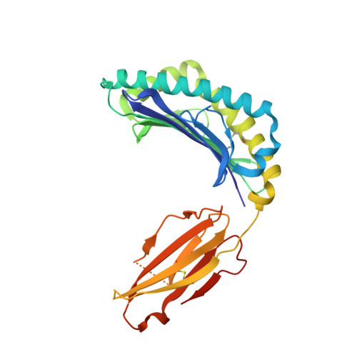
Peptide Centric V beta Specific Germline Contacts Shape a Specialist T Cell Response.

Wang, Y.Tsitsiklis, A.Devoe, S.Gao, W.Chu, H.H.Zhang, Y.Li, W.Wong, W.K.Deane, C.M.Neau, D.Slansky, J.E.Thomas, P.G.Robey, E.A.Dai, S.

(2022) Front Immunol 13: 847092-847092

  • DOI: https://doi.org/10.3389/fimmu.2022.847092
  • Primary Citation Related Structures: 
    8D5N, 8D5P, 8D5Q

  • PubMed Abstract: 

    Certain CD8 T cell responses are particularly effective at controlling infection, as exemplified by elite control of HIV in individuals harboring HLA-B57. To understand the structural features that contribute to CD8 T cell elite control, we focused on a strongly protective CD8 T cell response directed against a parasite-derived peptide (HF10) presented by an atypical MHC-I molecule, H-2Ld. This response exhibits a focused TCR repertoire dominated by Vβ2, and a representative TCR (TG6) in complex with Ld-HF10 reveals an unusual structure in which both MHC and TCR contribute extensively to peptide specificity, along with a parallel footprint of TCR on its pMHC ligand. The parallel footprint is a common feature of Vβ2-containing TCRs and correlates with an unusual Vα-Vβ interface, CDR loop conformations, and Vβ2-specific germline contacts with peptides. Vβ2 and Ld may represent "specialist" components for antigen recognition that allows for particularly strong and focused T cell responses.


  • Organizational Affiliation
    • Department of Pharmaceutical Sciences, University of Colorado School of Pharmacy, Aurora, CO, United States.

Macromolecule Content 

  • Total Structure Weight: 92.73 kDa 
  • Atom Count: 6,715 
  • Modeled Residue Count: 769 
  • Deposited Residue Count: 804 
  • Unique protein chains: 3

Macromolecules

Find similar proteins by:|  3D Structure
Entity ID: 1
MoleculeChains  Sequence LengthOrganismDetailsImage
H-2 class I histocompatibility antigen, L-D alpha chainA [auth C],
C [auth A]
282Mus musculusMutation(s): 0 
Gene Names: H2-L
UniProt
Find proteins for P01897 (Mus musculus)
Explore P01897 
Go to UniProtKB:  P01897
Entity Groups
Sequence Clusters30% Identity50% Identity70% Identity90% Identity95% Identity100% Identity
UniProt GroupP01897
Glycosylation
Glycosylation Sites: 1Go to GlyGen: P01897-1
Sequence Annotations
Expand
Reference Sequence
Find similar proteins by:  Sequence   |   3D Structure  
Entity ID: 2
MoleculeChains  Sequence LengthOrganismDetailsImage
Dense granule protein 6, HF10 peptideB [auth E],
D [auth B]
16Toxoplasma gondiiMutation(s): 0 
Gene Names: GRA6TG24
Find similar proteins by:|  3D Structure
Entity ID: 3
MoleculeChains  Sequence LengthOrganismDetailsImage
Beta-2-microglobulinE [auth F],
F [auth H]
104Mus musculusMutation(s): 0 
Gene Names: B2m
UniProt & NIH Common Fund Data Resources
Find proteins for P01887 (Mus musculus)
Explore P01887 
Go to UniProtKB:  P01887
IMPC:  MGI:88127
Entity Groups
Sequence Clusters30% Identity50% Identity70% Identity90% Identity95% Identity100% Identity
UniProt GroupP01887
Sequence Annotations
Expand
Reference Sequence

Small Molecules

Ligands 4 Unique
IDChains Name / Formula / InChI Key2D Diagram3D Interactions
NAG

Query on NAG


Download:Ideal Coordinates CCD File
G [auth C],
J [auth A]
2-acetamido-2-deoxy-beta-D-glucopyranose
C8 H15 N O6
OVRNDRQMDRJTHS-FMDGEEDCSA-N
PG4

Query on PG4


Download:Ideal Coordinates CCD File
H [auth C]TETRAETHYLENE GLYCOL
C8 H18 O5
UWHCKJMYHZGTIT-UHFFFAOYSA-N
PPI

Query on PPI


Download:Ideal Coordinates CCD File
I [auth E]PROPANOIC ACID
C3 H6 O2
XBDQKXXYIPTUBI-UHFFFAOYSA-N
EDO

Query on EDO


Download:Ideal Coordinates CCD File
K [auth F],
L [auth H]
1,2-ETHANEDIOL
C2 H6 O2
LYCAIKOWRPUZTN-UHFFFAOYSA-N

Experimental Data & Validation

Experimental Data

  • Method: X-RAY DIFFRACTION
  • Resolution: 1.80 Å
  • R-Value Free:  0.219 (Depositor), 0.223 (DCC) 
  • R-Value Work:  0.178 (Depositor), 0.186 (DCC) 
Space Group: C 1 2 1
Unit Cell:
Length ( Å )Angle ( ˚ )
a = 121.503α = 90
b = 139.816β = 130.824
c = 87.344γ = 90
Software Package:
Software NamePurpose
REFMACrefinement
XDSdata reduction
XDSdata scaling
PHASERphasing

Structure Validation

View Full Validation Report


Ligand Structure Quality Assessment 


Entry History 

& Funding Information

Deposition Data


Funding OrganizationLocationGrant Number
National Institutes of Health/National Institute Of Allergy and Infectious Diseases (NIH/NIAID)United States--

Revision History  (Full details and data files)

  • Version 1.0: 2022-09-14
    Type: Initial release
  • Version 1.1: 2023-10-18
    Changes: Data collection, Refinement description
  • Version 1.2: 2024-10-23
    Changes: Structure summary