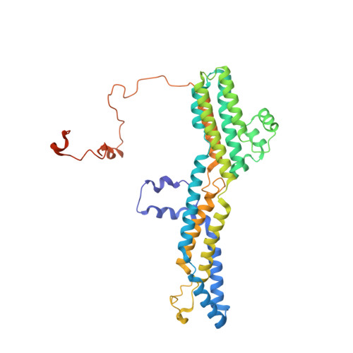
8D1F

hBest2 5mM Ca2+ (Ca2+-bound) closed state


Experimental Data Snapshot

  • Method: ELECTRON MICROSCOPY
  • Resolution: 1.82 Å
  • Aggregation State: PARTICLE 
  • Reconstruction Method: SINGLE PARTICLE 

wwPDB Validation   3D Report Full Report


This is version 1.1 of the entry. See complete history


Literature

Structures and gating mechanisms of human bestrophin anion channels.

Owji, A.P.Wang, J.Kittredge, A.Clark, Z.Zhang, Y.Hendrickson, W.A.Yang, T.

(2022) Nat Commun 13: 3836-3836

  • DOI: https://doi.org/10.1038/s41467-022-31437-7
  • Primary Citation of Related Structures:  
    8D1E, 8D1F, 8D1G, 8D1H, 8D1I, 8D1J, 8D1K, 8D1L, 8D1M, 8D1N, 8D1O

  • PubMed Abstract: 

    Bestrophin-1 (Best1) and bestrophin-2 (Best2) are two members of the bestrophin family of calcium (Ca 2+ )-activated chloride (Cl - ) channels with critical involvement in ocular physiology and direct pathological relevance. Here, we report cryo-EM structures of wild-type human Best1 and Best2 in various states at up to 1.8 Å resolution. Ca 2+ -bound Best1 structures illustrate partially open conformations at the two Ca 2+ -dependent gates of the channels, in contrast to the fully open conformations observed in Ca 2+ -bound Best2, which is in accord with the significantly smaller currents conducted by Best1 in electrophysiological recordings. Comparison of the closed and open states reveals a C-terminal auto-inhibitory segment (AS), which constricts the channel concentrically by wrapping around the channel periphery in an inter-protomer manner and must be released to allow channel opening. Our results demonstrate that removing the AS from Best1 and Best2 results in truncation mutants with similar activities, while swapping the AS between Best1 and Best2 results in chimeric mutants with swapped activities, underlying a key role of the AS in determining paralog specificity among bestrophins.


  • Organizational Affiliation

    Department of Ophthalmology, Columbia University, New York, NY, USA.


Macromolecules
Find similar proteins by:  (by identity cutoff)  |  3D Structure
Entity ID: 1
MoleculeChains Sequence LengthOrganismDetailsImage
Bestrophin-2A [auth C],
B [auth E],
C [auth A],
D,
E [auth B]
406Homo sapiensMutation(s): 0 
Gene Names: BEST2VMD2L1
Membrane Entity: Yes 
UniProt & NIH Common Fund Data Resources
Find proteins for Q8NFU1 (Homo sapiens)
Explore Q8NFU1 
Go to UniProtKB:  Q8NFU1
PHAROS:  Q8NFU1
GTEx:  ENSG00000039987 
Entity Groups  
Sequence Clusters30% Identity50% Identity70% Identity90% Identity95% Identity100% Identity
UniProt GroupQ8NFU1
Sequence Annotations
Expand
  • Reference Sequence
Small Molecules
Ligands 4 Unique
IDChains Name / Formula / InChI Key2D Diagram3D Interactions
MC3
Query on MC3

Download Ideal Coordinates CCD File 
AA [auth E]
AB [auth D]
BA [auth E]
BB [auth D]
CA [auth A]
AA [auth E],
AB [auth D],
BA [auth E],
BB [auth D],
CA [auth A],
EB [auth B],
FA [auth A],
FB [auth B],
GA [auth A],
GB [auth B],
H [auth C],
HA [auth A],
HB [auth B],
I [auth C],
IA [auth A],
IB [auth B],
J [auth C],
JA [auth A],
JB [auth B],
K [auth C],
KA [auth A],
KB [auth B],
L [auth C],
LA [auth A],
LB [auth B],
M [auth C],
MA [auth A],
MB [auth B],
N [auth C],
NA [auth A],
NB [auth B],
O [auth C],
OA [auth A],
P [auth C],
Q [auth C],
SA [auth D],
T [auth E],
TA [auth D],
U [auth E],
UA [auth D],
V [auth E],
VA [auth D],
W [auth E],
WA [auth D],
X [auth E],
XA [auth D],
Y [auth E],
YA [auth D],
Z [auth E],
ZA [auth D]
1,2-DIMYRISTOYL-RAC-GLYCERO-3-PHOSPHOCHOLINE
C36 H72 N O8 P
CITHEXJVPOWHKC-UUWRZZSWSA-N
DU0
Query on DU0

Download Ideal Coordinates CCD File 
DB [auth B],
EA [auth A],
G [auth C],
RA [auth D],
S [auth E]
2-[2-[(1~{S},2~{S},4~{S},5'~{R},6~{R},7~{S},8~{R},9~{S},12~{S},13~{R},16~{S})-5',7,9,13-tetramethylspiro[5-oxapentacyclo[10.8.0.0^{2,9}.0^{4,8}.0^{13,18}]icos-18-ene-6,2'-oxane]-16-yl]oxyethyl]propane-1,3-diol
C32 H52 O5
GFDJQXOBWHMOSQ-LEZUHYJESA-N
CA (Subject of Investigation/LOI)
Query on CA

Download Ideal Coordinates CCD File 
CB [auth B],
DA [auth A],
F [auth C],
QA [auth D],
R [auth E]
CALCIUM ION
Ca
BHPQYMZQTOCNFJ-UHFFFAOYSA-N
CL (Subject of Investigation/LOI)
Query on CL

Download Ideal Coordinates CCD File 
PA [auth A]CHLORIDE ION
Cl
VEXZGXHMUGYJMC-UHFFFAOYSA-M
Experimental Data & Validation

Experimental Data

  • Method: ELECTRON MICROSCOPY
  • Resolution: 1.82 Å
  • Aggregation State: PARTICLE 
  • Reconstruction Method: SINGLE PARTICLE 
EM Software:
TaskSoftware PackageVersion
RECONSTRUCTIONcryoSPARC
MODEL REFINEMENTPHENIX
MODEL REFINEMENTCoot
MODEL REFINEMENTREFMAC

Structure Validation

View Full Validation Report


Entry History & Funding Information

Deposition Data


Funding OrganizationLocationGrant Number
National Institutes of Health/National Eye Institute (NIH/NEI)United StatesEY030763-03
National Institutes of Health/National Institute of General Medical Sciences (NIH/NIGMS)United StatesGM107462-08
National Institutes of Health/National Eye Institute (NIH/NEI)United StatesGM127652-06

Revision History  (Full details and data files)

  • Version 1.0: 2022-07-13
    Type: Initial release
  • Version 1.1: 2024-02-14
    Changes: Data collection