8D1B | pdb_00008d1b

CryoEM structure of human orphan GPCR GPR179 in complex with extracellular matrix protein pikachurin


Experimental Data Snapshot

  • Method: ELECTRON MICROSCOPY
  • Resolution: 3.57 Å
  • Aggregation State: PARTICLE 
  • Reconstruction Method: SINGLE PARTICLE 

wwPDB Validation 3D Report Full Report

Validation slider image for 8D1B

This is version 1.2 of the entry. See complete history

Literature

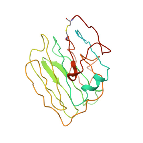
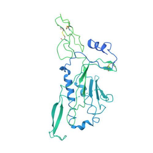
Structure of the photoreceptor synaptic assembly of the extracellular matrix protein pikachurin with the orphan receptor GPR179.

Patil, D.N.Pantalone, S.Cao, Y.Laboute, T.Novick, S.J.Singh, S.Savino, S.Faravelli, S.Magnani, F.Griffin, P.R.Singh, A.K.Forneris, F.Martemyanov, K.A.

(2023) Sci Signal 16: eadd9539-eadd9539

  • DOI: https://doi.org/10.1126/scisignal.add9539
  • Primary Citation Related Structures: 
    7ZC9, 7ZCB, 8D1B

  • PubMed Abstract: 

    Precise synapse formation is essential for normal functioning of the nervous system. Retinal photoreceptors establish selective contacts with bipolar cells, aligning the neurotransmitter release apparatus with postsynaptic signaling cascades. This involves transsynaptic assembly between the dystroglycan-dystrophin complex on the photoreceptor and the orphan receptor GPR179 on the bipolar cell, which is mediated by the extracellular matrix protein pikachurin (also known as EGFLAM). This complex plays a critical role in the synaptic organization of photoreceptors and signal transmission, and mutations affecting its components cause blinding disorders in humans. Here, we investigated the structural organization and molecular mechanisms by which pikachurin orchestrates transsynaptic assembly and solved structures of the human pikachurin domains by x-ray crystallography and of the GPR179-pikachurin complex by single-particle, cryo-electron microscopy. The structures reveal molecular recognition principles of pikachurin by the Cache domains of GPR179 and show how the interaction is involved in the transsynaptic alignment of the signaling machinery. Together, these data provide a structural basis for understanding the synaptic organization of photoreceptors and ocular pathology.


  • Organizational Affiliation
    • Department of Neuroscience, Herbert Wertheim UF Scripps Institute for Biomedical Innovation and Technology, University of Florida, Jupiter, FL 33458, USA.

Macromolecule Content 

  • Total Structure Weight: 214.86 kDa 
  • Atom Count: 6,724 
  • Modeled Residue Count: 874 
  • Deposited Residue Count: 1,948 
  • Unique protein chains: 2

Macromolecules

Find similar proteins by:|  3D Structure
Entity ID: 1
MoleculeChains  Sequence LengthOrganismDetailsImage
PikachurinA [auth D],
B [auth C]
234Homo sapiensMutation(s): 0 
Gene Names: EGFLAMAGRINLAGRNLPIKA
UniProt & NIH Common Fund Data Resources
Find proteins for Q63HQ2 (Homo sapiens)
Explore Q63HQ2 
Go to UniProtKB:  Q63HQ2
PHAROS:  Q63HQ2
GTEx:  ENSG00000164318 
Entity Groups
Sequence Clusters30% Identity50% Identity70% Identity90% Identity95% Identity100% Identity
UniProt GroupQ63HQ2
Sequence Annotations
Expand
Reference Sequence
Find similar proteins by:|  3D Structure
Entity ID: 2
MoleculeChains  Sequence LengthOrganismDetailsImage
Probable G-protein coupled receptor 179C [auth B],
D [auth A]
740Homo sapiensMutation(s): 0 
Gene Names: GPR179GPR158LGPR158L1
UniProt & NIH Common Fund Data Resources
Find proteins for Q6PRD1 (Homo sapiens)
Explore Q6PRD1 
Go to UniProtKB:  Q6PRD1
PHAROS:  Q6PRD1
Entity Groups
Sequence Clusters30% Identity50% Identity70% Identity90% Identity95% Identity100% Identity
UniProt GroupQ6PRD1
Sequence Annotations
Expand
Reference Sequence

Experimental Data & Validation

Experimental Data

  • Method: ELECTRON MICROSCOPY
  • Resolution: 3.57 Å
  • Aggregation State: PARTICLE 
  • Reconstruction Method: SINGLE PARTICLE 

Structure Validation

View Full Validation Report


Entry History 

& Funding Information

Deposition Data


Funding OrganizationLocationGrant Number
National Institutes of Health/National Institute of Mental Health (NIH/NIMH)United States--

Revision History  (Full details and data files)

  • Version 1.0: 2023-07-26
    Type: Initial release
  • Version 1.1: 2023-08-16
    Changes: Data collection, Database references
  • Version 1.2: 2024-10-09
    Changes: Data collection, Structure summary