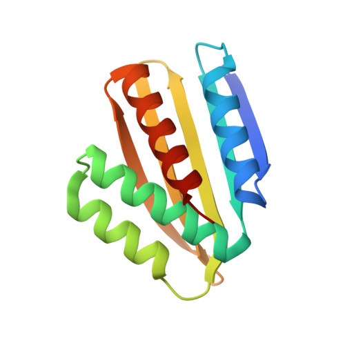
Robust deep learning-based protein sequence design using ProteinMPNN.
Dauparas, J., Anishchenko, I., Bennett, N., Bai, H., Ragotte, R.J., Milles, L.F., Wicky, B.I.M., Courbet, A., de Haas, R.J., Bethel, N., Leung, P.J.Y., Huddy, T.F., Pellock, S., Tischer, D., Chan, F., Koepnick, B., Nguyen, H., Kang, A., Sankaran, B., Bera, A.K., King, N.P., Baker, D.(2022) Science 378: 49-56
- PubMed: 36108050
- DOI: https://doi.org/10.1126/science.add2187
- Primary Citation of Related Structures:
8CYK - PubMed Abstract:
Although deep learning has revolutionized protein structure prediction, almost all experimentally characterized de novo protein designs have been generated using physically based approaches such as Rosetta. Here, we describe a deep learning-based protein sequence design method, ProteinMPNN, that has outstanding performance in both in silico and experimental tests. On native protein backbones, ProteinMPNN has a sequence recovery of 52.4% compared with 32.9% for Rosetta. The amino acid sequence at different positions can be coupled between single or multiple chains, enabling application to a wide range of current protein design challenges. We demonstrate the broad utility and high accuracy of ProteinMPNN using x-ray crystallography, cryo-electron microscopy, and functional studies by rescuing previously failed designs, which were made using Rosetta or AlphaFold, of protein monomers, cyclic homo-oligomers, tetrahedral nanoparticles, and target-binding proteins.
Organizational Affiliation:
Department of Biochemistry, University of Washington, Seattle, WA, USA.


