8CMM

Re-pairing DNA - binding of a ruthenium phi complex to a double mismatch

  • Classification: DNA
  • Organism(s): synthetic construct
  • Mutation(s): No 

  • Deposited: 2023-02-20 Released: 2024-03-06 
  • Deposition Author(s): Prieto Otoya, T.D., Cardin, C.J., McQuaid, K.M.
  • Funding Organization(s): H2020 Marie Curie Actions of the European Commission, Biotechnology and Biological Sciences Research Council (BBSRC)

Experimental Data Snapshot

  • Method: X-RAY DIFFRACTION
  • Resolution: 0.90 Å
  • R-Value Free: 0.159 
  • R-Value Work: 0.136 
  • R-Value Observed: 0.137 

wwPDB Validation   3D Report Full Report


Ligand Structure Quality Assessment 


This is version 2.1 of the entry. See complete history


Literature

Re-pairing DNA: binding of a ruthenium phi complex to a double mismatch.

Prieto Otoya, T.D.McQuaid, K.T.Paterson, N.G.Cardin, D.J.Kellett, A.Cardin, C.J.

(2024) Chem Sci 15: 9096-9103

  • DOI: https://doi.org/10.1039/d4sc01448k
  • Primary Citation of Related Structures:  
    8CMM

  • PubMed Abstract: 

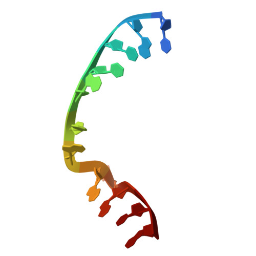
    We report a crystal structure at atomic resolution (0.9 Å) of a ruthenium complex bound to a consecutive DNA double mismatch, which results in a TA basepair with flipped out thymine, together with the formation of an adenine bulge. The structure shows a form of metalloinsertion interaction of the Λ-[Ru(phen) 2 phi] 2+ (phi = 9,10-phenanthrenediimine) complex at the bulge site. The metal complex interacts with the DNA via the major groove, where specific interactions between the adenines of the DNA and the phen ligands of the complex are formed. One Δ-[Ru(phen) 2 phi] 2+ complex interacts via the minor groove, which shows sandwiching of its phi ligand between the phi ligands of the other two ruthenium complexes, and no interaction of its phen ligands with DNA. To our knowledge, this binding model represents a new form of metalloinsertion in showing major rather than minor groove insertion.


  • Organizational Affiliation

    Department of Chemistry, University of Reading Whiteknights Reading, RG6 6AD UK c.j.cardin@reading.ac.uk.


Macromolecules

Find similar nucleic acids by:  Sequence   |   3D Structure  

Entity ID: 1
MoleculeChains LengthOrganismImage
DNA (5'-D(*CP*GP*CP*TP*AP*TP*AP*AP*TP*GP*CP*G)-3')12synthetic construct
Sequence Annotations
Expand
  • Reference Sequence
Experimental Data & Validation

Experimental Data

  • Method: X-RAY DIFFRACTION
  • Resolution: 0.90 Å
  • R-Value Free: 0.159 
  • R-Value Work: 0.136 
  • R-Value Observed: 0.137 
  • Space Group: I 21 21 21
Unit Cell:
Length ( Å )Angle ( ˚ )
a = 31.395α = 90
b = 48.645β = 90
c = 54.192γ = 90
Software Package:
Software NamePurpose
REFMACrefinement
DIALSdata scaling
DIALSdata reduction
SHELXphasing

Structure Validation

View Full Validation Report


Ligand Structure Quality Assessment 


Entry History & Funding Information

Deposition Data


Funding OrganizationLocationGrant Number
H2020 Marie Curie Actions of the European CommissionEuropean Union861381
Biotechnology and Biological Sciences Research Council (BBSRC)United KingdomBB/T008342/1

Revision History  (Full details and data files)

  • Version 1.0: 2024-03-06
    Type: Initial release
  • Version 2.0: 2024-06-12
    Type: Coordinate replacement
    Reason: Occupancy of atoms on special symmetry positions
    Changes: Advisory, Atomic model, Author supporting evidence, Data collection, Database references, Derived calculations, Non-polymer description, Other, Refinement description, Source and taxonomy, Structure summary
  • Version 2.1: 2024-07-03
    Changes: Database references