8C88 | pdb_00008c88

Double mutant G(M19)C/T(L214)C structure of Photosynthetic Reaction Center From Cereibacter sphaeroides strain RV


Experimental Data Snapshot

  • Method: X-RAY DIFFRACTION
  • Resolution: 2.75 Å
  • R-Value Free: 
    0.271 (Depositor), 0.247 (DCC) 
  • R-Value Work: 
    0.199 (Depositor) 
  • R-Value Observed: 
    0.202 (Depositor) 

Starting Model: experimental
View more details

wwPDB Validation 3D Report Full Report

Validation slider image for 8C88

Ligand Structure Quality Assessment 


This is version 1.1 of the entry. See complete history

Literature

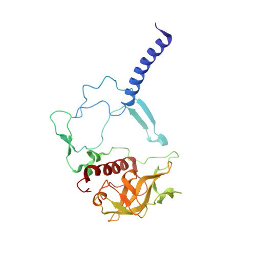
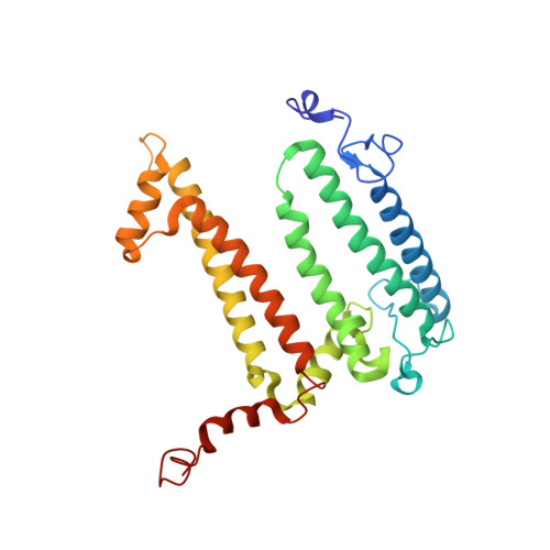
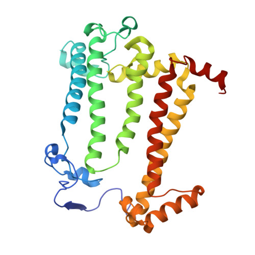
Stabilization of Cereibacter sphaeroides Photosynthetic Reaction Center by the Introduction of Disulfide Bonds.

Selikhanov, G.Atamas, A.Yukhimchuk, D.Fufina, T.Vasilieva, L.Gabdulkhakov, A.

(2023) Membranes (Basel) 13

  • DOI: https://doi.org/10.3390/membranes13020154
  • Primary Citation Related Structures: 
    8C5X, 8C6K, 8C7C, 8C87, 8C88

  • PubMed Abstract: 

    The photosynthetic reaction center of the purple nonsulfur bacterium Cereibacter sphaeroides is a useful model for the study of mechanisms of photoinduced electron transfer and a promising component for photo-bio-electrocatalytic systems. The basic research and technological applications of this membrane pigment-protein complex require effective approaches to increase its structural stability. In this work, a rational design approach to genetically modify the reaction centers by introducing disulfide bonds is used. This resulted in significantly increasing the thermal stability of some of the mutant pigment-protein complexes. The formation of the S-S bonds was confirmed by X-ray crystallography as well as SDS-PAGE, and the optical properties of the reaction centers were studied. The genetically modified reaction centers presented here preserved their ability for photochemical charge separation and could be of interest for basic science and biotechnology.


  • Organizational Affiliation
    • Institute of Protein Research, Russian Academy of Sciences, Institutskaya 4, 142290 Pushchino, Moscow Region, Russia.

Macromolecule Content 

  • Total Structure Weight: 101.72 kDa 
  • Atom Count: 7,341 
  • Modeled Residue Count: 826 
  • Deposited Residue Count: 826 
  • Unique protein chains: 3

Macromolecules

Find similar proteins by:|  3D Structure
Entity ID: 1
MoleculeChains  Sequence LengthOrganismDetailsImage
Reaction center protein H chainA [auth H]242Cereibacter sphaeroides 2.4.1Mutation(s): 0 
Gene Names: puhA
Membrane Entity: Yes 
UniProt
Find proteins for Q3J170 (Cereibacter sphaeroides (strain ATCC 17023 / DSM 158 / JCM 6121 / CCUG 31486 / LMG 2827 / NBRC 12203 / NCIMB 8253 / ATH 2.4.1.))
Explore Q3J170 
Go to UniProtKB:  Q3J170
Entity Groups
Sequence Clusters30% Identity50% Identity70% Identity90% Identity95% Identity100% Identity
UniProt GroupQ3J170
Sequence Annotations
Expand
Reference Sequence
Find similar proteins by:|  3D Structure
Entity ID: 2
MoleculeChains  Sequence LengthOrganismDetailsImage
Reaction center protein L chainB [auth L]281Cereibacter sphaeroides 2.4.1Mutation(s): 2 
Gene Names: pufL
Membrane Entity: Yes 
UniProt
Find proteins for Q3J1A5 (Cereibacter sphaeroides (strain ATCC 17023 / DSM 158 / JCM 6121 / CCUG 31486 / LMG 2827 / NBRC 12203 / NCIMB 8253 / ATH 2.4.1.))
Explore Q3J1A5 
Go to UniProtKB:  Q3J1A5
Entity Groups
Sequence Clusters30% Identity50% Identity70% Identity90% Identity95% Identity100% Identity
UniProt GroupQ3J1A5
Sequence Annotations
Expand
Reference Sequence
Find similar proteins by:|  3D Structure
Entity ID: 3
MoleculeChains  Sequence LengthOrganismDetailsImage
Reaction center protein M chainC [auth M]303Cereibacter sphaeroides 2.4.1Mutation(s): 2 
Gene Names: pufM
Membrane Entity: Yes 
UniProt
Find proteins for Q3J1A6 (Cereibacter sphaeroides (strain ATCC 17023 / DSM 158 / JCM 6121 / CCUG 31486 / LMG 2827 / NBRC 12203 / NCIMB 8253 / ATH 2.4.1.))
Explore Q3J1A6 
Go to UniProtKB:  Q3J1A6
Entity Groups
Sequence Clusters30% Identity50% Identity70% Identity90% Identity95% Identity100% Identity
UniProt GroupQ3J1A6
Sequence Annotations
Expand
Reference Sequence

Small Molecules

Ligands 12 Unique
IDChains Name / Formula / InChI Key2D Diagram3D Interactions
BCL
(Subject of Investigation/LOI)

Query on BCL


Download:Ideal Coordinates CCD File
J [auth L],
K [auth L],
U [auth M],
V [auth M]
BACTERIOCHLOROPHYLL A
C55 H74 Mg N4 O6
DSJXIQQMORJERS-AGGZHOMASA-M
BPH
(Subject of Investigation/LOI)

Query on BPH


Download:Ideal Coordinates CCD File
L,
W [auth M]
BACTERIOPHEOPHYTIN A
C55 H76 N4 O6
KWOZSBGNAHVCKG-SZQBJALDSA-N
U10
(Subject of Investigation/LOI)

Query on U10


Download:Ideal Coordinates CCD File
Y [auth M]UBIQUINONE-10
C59 H90 O4
ACTIUHUUMQJHFO-UPTCCGCDSA-N
SPN
(Subject of Investigation/LOI)

Query on SPN


Download:Ideal Coordinates CCD File
Z [auth M]SPEROIDENONE
C41 H70 O2
GWQAMGYOEYXWJF-YCDPMLDASA-N
NKP

Query on NKP


Download:Ideal Coordinates CCD File
D [auth H],
T [auth M]
(2R)-2-hydroxy-3-(phosphonooxy)propyl (9E)-octadec-9-enoate
C21 H41 O7 P
WRGQSWVCFNIUNZ-SQUSKLHYSA-N
OLC
(Subject of Investigation/LOI)

Query on OLC


Download:Ideal Coordinates CCD File
I [auth L](2R)-2,3-dihydroxypropyl (9Z)-octadec-9-enoate
C21 H40 O4
RZRNAYUHWVFMIP-GDCKJWNLSA-N
LDA

Query on LDA


Download:Ideal Coordinates CCD File
AA [auth M]
BA [auth M]
CA [auth M]
DA [auth M]
E [auth H]
AA [auth M],
BA [auth M],
CA [auth M],
DA [auth M],
E [auth H],
HA [auth M]
LAURYL DIMETHYLAMINE-N-OXIDE
C14 H31 N O
SYELZBGXAIXKHU-UHFFFAOYSA-N
PO4

Query on PO4


Download:Ideal Coordinates CCD File
FA [auth M],
GA [auth M]
PHOSPHATE ION
O4 P
NBIIXXVUZAFLBC-UHFFFAOYSA-K
DIO

Query on DIO


Download:Ideal Coordinates CCD File
P [auth L]1,4-DIETHYLENE DIOXIDE
C4 H8 O2
RYHBNJHYFVUHQT-UHFFFAOYSA-N
EDO

Query on EDO


Download:Ideal Coordinates CCD File
Q [auth L],
R [auth L],
S [auth L]
1,2-ETHANEDIOL
C2 H6 O2
LYCAIKOWRPUZTN-UHFFFAOYSA-N
FE
(Subject of Investigation/LOI)

Query on FE


Download:Ideal Coordinates CCD File
X [auth M]FE (III) ION
Fe
VTLYFUHAOXGGBS-UHFFFAOYSA-N
UNL

Query on UNL


Download:Ideal Coordinates CCD File
EA [auth M]
F [auth H]
G [auth H]
H
M [auth L]
EA [auth M],
F [auth H],
G [auth H],
H,
M [auth L],
N [auth L],
O [auth L]
Unknown ligand
VTLYFUHAOXGGBS-UHFFFAOYSA-N

Experimental Data & Validation

Experimental Data

  • Method: X-RAY DIFFRACTION
  • Resolution: 2.75 Å
  • R-Value Free:  0.271 (Depositor), 0.247 (DCC) 
  • R-Value Work:  0.199 (Depositor) 
  • R-Value Observed: 0.202 (Depositor) 
Space Group: P 42 21 2
Unit Cell:
Length ( Å )Angle ( ˚ )
a = 100.91α = 90
b = 100.91β = 90
c = 237.02γ = 90
Software Package:
Software NamePurpose
REFMACrefinement
PHENIXrefinement
XDSdata reduction
XSCALEdata scaling
PHASERphasing

Structure Validation

View Full Validation Report


Ligand Structure Quality Assessment 


Entry History 

& Funding Information

Deposition Data


Funding OrganizationLocationGrant Number
Russian Foundation for Basic ResearchRussian Federation18-02-40008

Revision History  (Full details and data files)

  • Version 1.0: 2023-11-22
    Type: Initial release
  • Version 1.1: 2024-10-16
    Changes: Structure summary