8C7R | pdb_00008c7r

Crystal structure of rat autotaxin and compound MEY-003


Experimental Data Snapshot

  • Method: X-RAY DIFFRACTION
  • Resolution: 2.53 Å
  • R-Value Free: 
    0.254 (Depositor), 0.254 (DCC) 
  • R-Value Work: 
    0.205 (Depositor), 0.205 (DCC) 
  • R-Value Observed: 
    0.209 (Depositor) 

Starting Model: experimental
View more details

wwPDB Validation 3D Report Full Report

Validation slider image for 8C7R

Ligand Structure Quality Assessment 


This is version 1.2 of the entry. See complete history

Literature

Discovery of potent chromone-based autotaxin inhibitors inspired by cannabinoids.

Eymery, M.C.Nguyen, K.A.Basu, S.Hausmann, J.Tran-Nguyen, V.K.Seidel, H.P.Gutierrez, L.Boumendjel, A.McCarthy, A.A.

(2023) Eur J Med Chem 263: 115944-115944

  • DOI: https://doi.org/10.1016/j.ejmech.2023.115944
  • Primary Citation Related Structures: 
    8C3O, 8C3P, 8C4W, 8C7R

  • PubMed Abstract: 

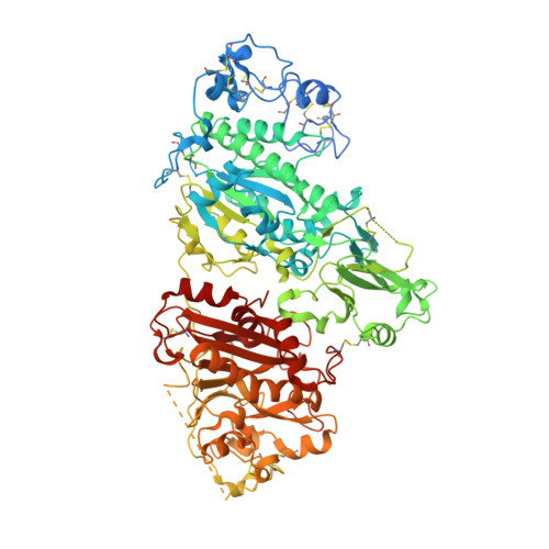
    Autotaxin (ATX) is an enzyme primarily known for the production of lysophosphatidic acid. Being involved in the development of major human diseases, such as cancer and neurodegenerative diseases, the enzyme has been featured in multiple studies as a pharmacological target. We previously found that the cannabinoid tetrahydrocannabinol (THC) could bind and act as an excellent inhibitor of ATX. This study aims to use the cannabinoid scaffold as a starting point to find cannabinoid-unrelated ATX inhibitors, following a funnel down approach in which large chemical libraries sharing chemical similarities with THC were screened to identify lead scaffold types for optimization. This approach allowed us to identify compounds bearing chromone and indole scaffolds as promising ATX inhibitors. Further optimization led to MEY-003, which is characterized by the direct linkage of an N-pentyl indole to the 5,7-dihydroxychromone moiety. This molecule has potent inhibitory activity towards ATX-β and ATX-ɣ as evidenced by enzymatic studies and its mode of action was rationalized by structural biology studies using macromolecular X-ray crystallography.


  • Organizational Affiliation
    • European Molecular Biology Laboratory, EMBL Grenoble, 71 Avenue des Martyrs, 38000, Grenoble, France; Univ. Grenoble Alpes, INSERM U1039, LRB, 38000, Grenoble, France.

Macromolecule Content 

  • Total Structure Weight: 100.74 kDa 
  • Atom Count: 6,448 
  • Modeled Residue Count: 775 
  • Deposited Residue Count: 862 
  • Unique protein chains: 1

Macromolecules

Find similar proteins by:|  3D Structure
Entity ID: 1
MoleculeChains  Sequence LengthOrganismDetailsImage
Ectonucleotide pyrophosphatase/phosphodiesterase family member 2862Rattus norvegicusMutation(s): 2 
Gene Names: Enpp2AtxNpps2
EC: 3.1.4.39 (PDB Primary Data), 3.1.4.4 (UniProt)
UniProt
Find proteins for Q64610 (Rattus norvegicus)
Explore Q64610 
Go to UniProtKB:  Q64610
Entity Groups
Sequence Clusters30% Identity50% Identity70% Identity90% Identity95% Identity100% Identity
UniProt GroupQ64610
Glycosylation
Glycosylation Sites: 1Go to GlyGen: Q64610-1
Sequence Annotations
Expand
Reference Sequence

Oligosaccharides

Help  
Entity ID: 2
MoleculeChains Length2D Diagram GlycosylationD Interactions
2-acetamido-2-deoxy-beta-D-glucopyranose-(1-4)-2-acetamido-2-deoxy-beta-D-glucopyranose
B
2N-Glycosylation
Glycosylation Resources
GlyTouCan: G42666HT
GlyCosmos: G42666HT
GlyGen: G42666HT

Small Molecules

Ligands 5 Unique
IDChains Name / Formula / InChI Key2D Diagram3D Interactions
5JK

Query on 5JK


Download:Ideal Coordinates CCD File
D [auth A]7alpha-hydroxycholesterol
C27 H46 O2
OYXZMSRRJOYLLO-RVOWOUOISA-N
T8R
(Subject of Investigation/LOI)

Query on T8R


Download:Ideal Coordinates CCD File
C [auth A]5,7-bis(oxidanyl)-2-(1-pentylindol-3-yl)chromen-4-one
C22 H21 N O4
RZGZFONKKPMOLH-UHFFFAOYSA-N
IOD

Query on IOD


Download:Ideal Coordinates CCD File
G [auth A],
H [auth A],
I [auth A],
J [auth A]
IODIDE ION
I
XMBWDFGMSWQBCA-UHFFFAOYSA-M
ZN

Query on ZN


Download:Ideal Coordinates CCD File
E [auth A],
F [auth A]
ZINC ION
Zn
PTFCDOFLOPIGGS-UHFFFAOYSA-N
CA

Query on CA


Download:Ideal Coordinates CCD File
K [auth A],
L [auth A]
CALCIUM ION
Ca
BHPQYMZQTOCNFJ-UHFFFAOYSA-N
Binding Affinity Annotations 
IDSourceBinding Affinity
T8R BindingDB:  8C7R Ki: 432 (nM) from 1 assay(s)
IC50: min: 380, max: 1099 (nM) from 4 assay(s)

Experimental Data & Validation

Experimental Data

  • Method: X-RAY DIFFRACTION
  • Resolution: 2.53 Å
  • R-Value Free:  0.254 (Depositor), 0.254 (DCC) 
  • R-Value Work:  0.205 (Depositor), 0.205 (DCC) 
  • R-Value Observed: 0.209 (Depositor) 
Space Group: P 1
Unit Cell:
Length ( Å )Angle ( ˚ )
a = 53.529α = 104.01
b = 61.843β = 98.43
c = 63.85γ = 93.27
Software Package:
Software NamePurpose
PHENIXrefinement
XDSdata reduction
Aimlessdata scaling
PHASERphasing
autoPROCdata processing

Structure Validation

View Full Validation Report


Ligand Structure Quality Assessment 


Entry History 

& Funding Information

Deposition Data


Funding OrganizationLocationGrant Number
European Molecular Biology Organization (EMBO)European Union--

Revision History  (Full details and data files)

  • Version 1.0: 2023-11-22
    Type: Initial release
  • Version 1.1: 2023-11-29
    Changes: Database references
  • Version 1.2: 2024-10-23
    Changes: Structure summary