


Structure and Dynamics of the Penicillin-Binding Protein 3 from Staphylococcus Epidermidis Native and in Complex with Cefotaxime and Vaborbactam
Schwinzer, M., Brognaro, H., Rohde, H., Betzel, C.(2023) Int J Appl Biol Pharm
Experimental Data Snapshot
Starting Model: experimental
View more details
(2023) Int J Appl Biol Pharm
Entity ID: 1 | |||||
|---|---|---|---|---|---|
| Molecule | Chains | Sequence Length | Organism | Details | Image |
| Penicillin-binding protein 3 | 668 | Staphylococcus epidermidis RP62A | Mutation(s): 0 Gene Names: pbp3, SERP1117 | 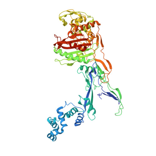 | |
UniProt | |||||
Entity Groups | |||||
| Sequence Clusters | 30% Identity50% Identity70% Identity90% Identity95% Identity100% Identity | ||||
| UniProt Group | Q5HNZ7 | ||||
Sequence AnnotationsExpand | |||||
Reference Sequence | |||||
| Ligands 1 Unique | |||||
|---|---|---|---|---|---|
| ID | Chains | Name / Formula / InChI Key | 2D Diagram | 3D Interactions | |
| CEF (Subject of Investigation/LOI) Download:Ideal Coordinates CCD File | B [auth A] | CEFOTAXIME, C3' cleaved, open, bound form C14 H15 N5 O5 S2 NRYMPLKBKFIWQC-YVCCLBOHSA-N |  | ||
| Length ( Å ) | Angle ( ˚ ) |
|---|---|
| a = 83.061 | α = 90 |
| b = 83.061 | β = 90 |
| c = 308.864 | γ = 90 |
| Software Name | Purpose |
|---|---|
| PHENIX | refinement |
| PHENIX | refinement |
| XDS | data reduction |
| XSCALE | data scaling |
| PHENIX | phasing |
| Funding Organization | Location | Grant Number |
|---|---|---|
| German Research Foundation (DFG) | Germany | -- |


