8C4E | pdb_00008c4e

F-actin decorated by SipA426-685


Experimental Data Snapshot

  • Method: ELECTRON MICROSCOPY
  • Resolution: 2.60 Å
  • Aggregation State: FILAMENT 
  • Reconstruction Method: HELICAL 

wwPDB Validation 3D Report Full Report

Validation slider image for 8C4E

This is version 1.0 of the entry. See complete history

Literature

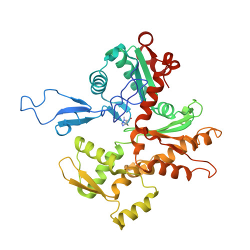
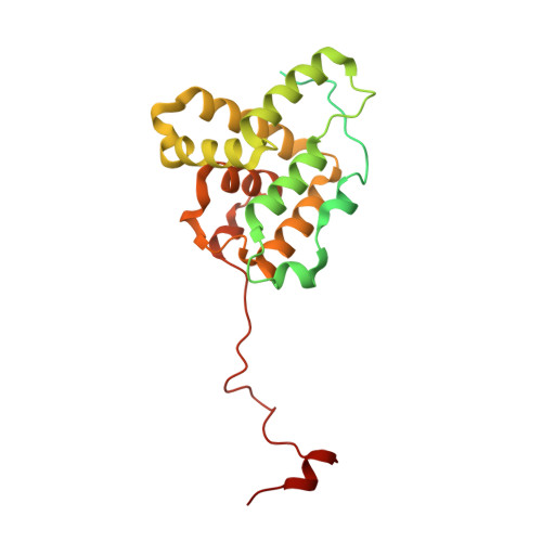
Structural basis for subversion of host cell actin cytoskeleton during Salmonella infection.

Yuan, B.Scholz, J.Wald, J.Thuenauer, R.Hennell James, R.Ellenberg, I.Windhorst, S.Faix, J.Marlovits, T.C.

(2023) Sci Adv 9: eadj5777-eadj5777

  • DOI: https://doi.org/10.1126/sciadv.adj5777
  • Primary Citation Related Structures: 
    8C4C, 8C4E

  • PubMed Abstract: 

    Secreted bacterial type III secretion system (T3SS) proteins are essential for successful infection by many human pathogens. Both T3SS translocator SipC and effector SipA are critical for Salmonella infection by subversion of the host cell cytoskeleton, but the precise molecular interplay between them remains unknown. Here, using cryo-electron microscopy, we show that SipA binds along the F-actin grooves with a unique binding pattern. SipA stabilizes F-actin through charged interface residues and appears to prevent inorganic phosphate release through closure of the "back door" of adenosine 5'-triphosphate pocket. We also show that SipC enhances the binding of SipA to F-actin, thus demonstrating that a sequential presence of T3SS proteins in host cells is associated with a sequence of infection events-starting with actin nucleation, filament growth, and stabilization. Together, our data explain the coordinated interplay of a precisely tuned and highly effective mechanism during Salmonella infection and provide a blueprint for interfering with Salmonella effectors acting on actin.


  • Organizational Affiliation
    • University Medical Center Hamburg-Eppendorf (UKE), Institute of Structural and Systems Biology, Hamburg, Germany.

Macromolecule Content 

  • Total Structure Weight: 454.91 kDa 
  • Atom Count: 28,796 
  • Modeled Residue Count: 3,648 
  • Deposited Residue Count: 4,060 
  • Unique protein chains: 2

Macromolecules

Find similar proteins by:|  3D Structure
Entity ID: 1
MoleculeChains  Sequence LengthOrganismDetailsImage
Actin, alpha skeletal muscle
A, B, C, D, E
A, B, C, D, E, F, G, H
377Gallus gallusMutation(s): 0 
EC: 3.6.4
UniProt
Find proteins for P68139 (Gallus gallus)
Explore P68139 
Go to UniProtKB:  P68139
Entity Groups
Sequence Clusters30% Identity50% Identity70% Identity90% Identity95% Identity100% Identity
UniProt GroupP68139
Sequence Annotations
Expand
Reference Sequence
Find similar proteins by:|  3D Structure
Entity ID: 2
MoleculeChains  Sequence LengthOrganismDetailsImage
Cell invasion protein SipAI [auth K],
J [auth L],
K [auth M],
L [auth N]
261SalmonellaMutation(s): 0 
Gene Names: sipAsspA
UniProt
Find proteins for P0CL52 (Salmonella typhimurium (strain LT2 / SGSC1412 / ATCC 700720))
Explore P0CL52 
Go to UniProtKB:  P0CL52
Entity Groups
Sequence Clusters30% Identity50% Identity70% Identity90% Identity95% Identity100% Identity
UniProt GroupP0CL52
Sequence Annotations
Expand
Reference Sequence

Small Molecules

Ligands 3 Unique
IDChains Name / Formula / InChI Key2D Diagram3D Interactions
ADP
(Subject of Investigation/LOI)

Query on ADP


Download:Ideal Coordinates CCD File
CA [auth F]
FA [auth G]
JA [auth H]
M [auth A]
Q [auth B]
CA [auth F],
FA [auth G],
JA [auth H],
M [auth A],
Q [auth B],
S [auth C],
W [auth D],
Z [auth E]
ADENOSINE-5'-DIPHOSPHATE
C10 H15 N5 O10 P2
XTWYTFMLZFPYCI-KQYNXXCUSA-N
PO4
(Subject of Investigation/LOI)

Query on PO4


Download:Ideal Coordinates CCD File
AA [auth E]
DA [auth F]
EA [auth G]
IA [auth H]
N [auth A]
AA [auth E],
DA [auth F],
EA [auth G],
IA [auth H],
N [auth A],
P [auth B],
U [auth C],
X [auth D]
PHOSPHATE ION
O4 P
NBIIXXVUZAFLBC-UHFFFAOYSA-K
MG
(Subject of Investigation/LOI)

Query on MG


Download:Ideal Coordinates CCD File
BA [auth F]
GA [auth G]
HA [auth H]
O [auth A]
R [auth B]
BA [auth F],
GA [auth G],
HA [auth H],
O [auth A],
R [auth B],
T [auth C],
V [auth D],
Y [auth E]
MAGNESIUM ION
Mg
JLVVSXFLKOJNIY-UHFFFAOYSA-N
Modified Residues  1 Unique
IDChains TypeFormula2D DiagramParent
HIC
Query on HIC
A, B, C, D, E
A, B, C, D, E, F, G, H
L-PEPTIDE LINKINGC7 H11 N3 O2HIS

Experimental Data & Validation

Experimental Data

  • Method: ELECTRON MICROSCOPY
  • Resolution: 2.60 Å
  • Aggregation State: FILAMENT 
  • Reconstruction Method: HELICAL 
EM Software:
TaskSoftware PackageVersion
RECONSTRUCTIONRELION3.1

Structure Validation

View Full Validation Report


Entry History 

& Funding Information

Deposition Data


Funding OrganizationLocationGrant Number
German Research Foundation (DFG)Germanynumbers INST152/772-1, 152/774-1, 152/775-1, 152/776-1 and 152/777-1 FUGG.

Revision History  (Full details and data files)

  • Version 1.0: 2024-01-17
    Type: Initial release