8BZ8 | pdb_00008bz8

Crystal structure of the L. monocytogenes RmlT D198A variant in complex with TDP-rhamnose


Experimental Data Snapshot

  • Method: X-RAY DIFFRACTION
  • Resolution: 2.52 Å
  • R-Value Free: 
    0.228 (Depositor), 0.227 (DCC) 
  • R-Value Work: 
    0.191 (Depositor), 0.191 (DCC) 
  • R-Value Observed: 
    0.193 (Depositor) 

Starting Model: experimental
View more details

wwPDB Validation 3D Report Full Report

Validation slider image for 8BZ8

Ligand Structure Quality Assessment 


This is version 1.2 of the entry. See complete history

Literature

Molecular properties of the RmlT wall teichoic acid rhamnosyltransferase that modulates virulence in Listeria monocytogenes.

Monteiro, R.Cereija, T.B.Pombinho, R.Voskuilen, T.Codee, J.D.C.Sousa, S.Morais-Cabral, J.H.Cabanes, D.

(2025) Nat Commun 16: 24-24

  • DOI: https://doi.org/10.1038/s41467-024-55360-1
  • Primary Citation Related Structures: 
    8BZ4, 8BZ5, 8BZ6, 8BZ7, 8BZ8, 9GZJ, 9GZK

  • PubMed Abstract: 

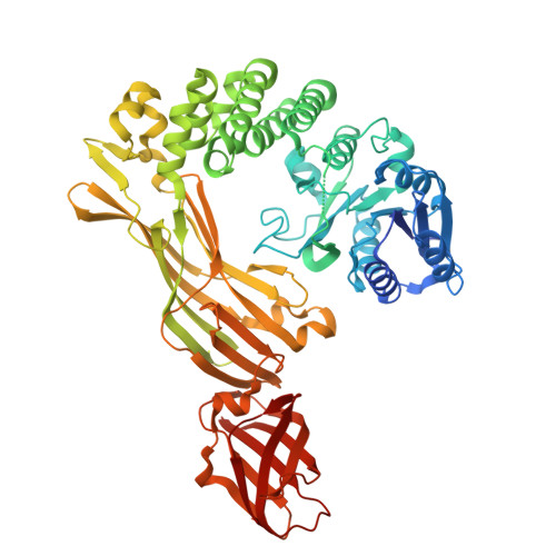
    Wall teichoic acids (WTAs) from the major Gram-positive foodborne pathogen Listeria monocytogenes are peptidoglycan-associated glycopolymers decorated by monosaccharides that, while not essential for bacterial growth, are required for bacterial virulence and resistance to antimicrobials. Here we report the structure and function of a bacterial WTAs rhamnosyltransferase, RmlT, strictly required for L. monocytogenes WTAs rhamnosylation. In particular, we demonstrated that RmlT transfers rhamnose from dTDP-L-rhamnose to naked WTAs, and that specificity towards TDP-rhamnose is not determined by its binding affinity. Structures of RmlT with and without its substrates showed that this enzyme is a dimer, revealed the residues responsible for interaction with the substrates and that the catalytic residue pre-orients the acceptor substrate towards the nucleophilic attack to the sugar. Additionally, the structures provided indications for two potential interaction pathways for the long WTAs on the surface of RmlT. Finally, we confirmed that WTAs glycosyltransferases are promising targets for next-generation strategies against Gram-positive pathogens by showing that inactivation of the RmlT catalytic activity results in a decreased infection in vivo.


  • Organizational Affiliation
    • i3S - Instituto de Investigação e Inovação em Saúde, Universidade do Porto, Porto, Portugal. ricardo.monteiro@i3s.up.pt.

Macromolecule Content 

  • Total Structure Weight: 433.89 kDa 
  • Atom Count: 30,233 
  • Modeled Residue Count: 3,608 
  • Deposited Residue Count: 3,744 
  • Unique protein chains: 1

Macromolecules

Find similar proteins by:|  3D Structure
Entity ID: 1
MoleculeChains  Sequence LengthOrganismDetailsImage
Glycosyltransferase
A, B, C, D, E
A, B, C, D, E, F
624Listeria monocytogenesMutation(s): 1 
Gene Names: APD94_03445
EC: 2.4
UniProt
Find proteins for A0A401AAP7 (Listeria monocytogenes)
Explore A0A401AAP7 
Go to UniProtKB:  A0A401AAP7
Entity Groups
Sequence Clusters30% Identity50% Identity70% Identity90% Identity95% Identity100% Identity
UniProt GroupA0A401AAP7
Sequence Annotations
Expand
Reference Sequence

Small Molecules

Ligands 4 Unique
IDChains Name / Formula / InChI Key2D Diagram3D Interactions
TRH
(Subject of Investigation/LOI)

Query on TRH


Download:Ideal Coordinates CCD File
I [auth A]
K [auth B]
P [auth C]
R [auth D]
V [auth E]
I [auth A],
K [auth B],
P [auth C],
R [auth D],
V [auth E],
Y [auth F]
2'-DEOXY-THYMIDINE-BETA-L-RHAMNOSE
C16 H26 N2 O15 P2
ZOSQFDVXNQFKBY-CGAXJHMRSA-N
TAR

Query on TAR


Download:Ideal Coordinates CCD File
AA [auth F],
BA [auth F],
M [auth B],
S [auth D],
W [auth E]
D(-)-TARTARIC ACID
C4 H6 O6
FEWJPZIEWOKRBE-LWMBPPNESA-N
CL

Query on CL


Download:Ideal Coordinates CCD File
L [auth B],
Z [auth F]
CHLORIDE ION
Cl
VEXZGXHMUGYJMC-UHFFFAOYSA-M
MG

Query on MG


Download:Ideal Coordinates CCD File
G [auth A]
H [auth A]
J [auth B]
N [auth C]
O [auth C]
G [auth A],
H [auth A],
J [auth B],
N [auth C],
O [auth C],
Q [auth D],
T [auth E],
U [auth E],
X [auth F]
MAGNESIUM ION
Mg
JLVVSXFLKOJNIY-UHFFFAOYSA-N

Experimental Data & Validation

Experimental Data

  • Method: X-RAY DIFFRACTION
  • Resolution: 2.52 Å
  • R-Value Free:  0.228 (Depositor), 0.227 (DCC) 
  • R-Value Work:  0.191 (Depositor), 0.191 (DCC) 
  • R-Value Observed: 0.193 (Depositor) 
Space Group: P 1 21 1
Unit Cell:
Length ( Å )Angle ( ˚ )
a = 87.397α = 90
b = 292.381β = 100.498
c = 93.075γ = 90
Software Package:
Software NamePurpose
PHENIXrefinement
XDSdata scaling
PHASERphasing
XDSdata reduction

Structure Validation

View Full Validation Report


Ligand Structure Quality Assessment 


Entry History 

& Funding Information

Deposition Data


Funding OrganizationLocationGrant Number
Fundacao para a Ciencia e a TecnologiaPortugal--

Revision History  (Full details and data files)

  • Version 1.0: 2023-12-27
    Type: Initial release
  • Version 1.1: 2025-01-15
    Changes: Database references, Structure summary
  • Version 1.2: 2025-02-05
    Changes: Refinement description