Structural insights into a cooperative switch between one and two FimH bacterial adhesins binding pauci- and high-mannose type N-glycan receptors.
Krammer, E.M., Bridot, C., Serna, S., Echeverria, B., Semwal, S., Roubinet, B., van Noort, K., Wilbers, R.H.P., Bourenkov, G., de Ruyck, J., Landemarre, L., Reichardt, N., Bouckaert, J.(2023) J Biological Chem 299: 104627-104627
- PubMed: 36944399 Search on PubMedSearch on PubMed Central
- DOI: https://doi.org/10.1016/j.jbc.2023.104627
- Primary Citation Related Structures:
7BHD, 8BXY, 8BY3 - PubMed Abstract:
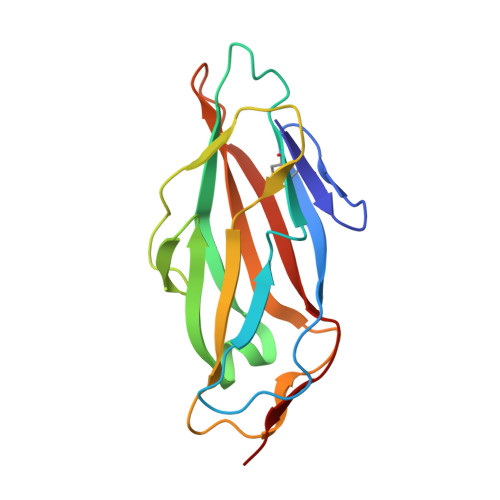
The FimH type-1 fimbrial adhesin allows pathogenic Escherichia coli to adhere to glycoproteins in the epithelial linings of human bladder and intestinal tract, by using multiple fimbriae simultaneously. Pauci- and high-mannose type N-glycans are natural FimH receptors on those glycoproteins. Oligomannose-3 and oligomannose-5 bind with the highest affinity to FimH by using the same Manα1,3Man branch. Oligomannose-6 is generated from oligomannose-5 in the next step of the biogenesis of high-mannose N-glycans, by the transfer of a mannose in α1,2-linkage onto this branch. Using serial crystallography and by measuring the kinetics of binding, we demonstrate that shielding the high-affinity epitope drives the binding of multiple FimH molecules. First, we profiled FimH glycan binding on a microarray containing paucimannosidic N-glycans and in a FimH LEctPROFILE assay. To make the transition to oligomannose-6, we measured the kinetics of FimH binding using paucimannosidic N-glycans, glycoproteins and all four α-dimannosides conjugated to bovine serum albumin. Equimolar mixed interfaces of the dimannosides present in oligomannose-6 and molecular dynamics simulations suggest a positive cooperativity in the bivalent binding of Manα1,3Manα1 and Manα1,6Manα1 dimannosides. The binding of core α1,6-fucosylated oligomannose-3 in cocrystals of FimH is monovalent but interestingly the GlcNAc1-Fuc moiety retains highly flexibility. In cocrystals with oligomannose-6, two FimH bacterial adhesins bind the Manα1,3Manα1 and Manα1,6Manα1 endings of the second trimannose core (A-4'-B). This cooperative switch towards bivalent binding appears sustainable beyond a molar excess of oligomannose-6. Our findings provide important novel structural insights for the design of multivalent FimH antagonists that bind with positive cooperativity.
- Unité de Glycobiologie Structurale et Fonctionnelle (UGSF), UMR 8576 CNRS and University of Lille, Villeneuve d'Ascq, France.
Organizational Affiliation: