8BVL | pdb_00008bvl

Crystal structure of the IBR-RING2 domain of HOIL-1


Experimental Data Snapshot

  • Method: X-RAY DIFFRACTION
  • Resolution: 2.24 Å
  • R-Value Free: 
    0.296 (Depositor), 0.297 (DCC) 
  • R-Value Work: 
    0.234 (Depositor) 
  • R-Value Observed: 
    0.237 (Depositor) 

Starting Model: experimental
View more details

wwPDB Validation 3D Report Full Report

Validation slider image for 8BVL

This is version 1.2 of the entry. See complete history

Literature

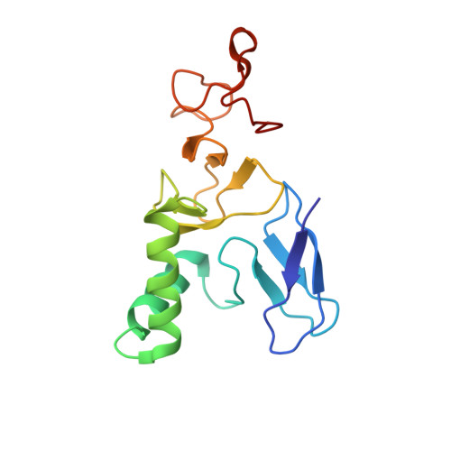
Structural basis for ubiquitylation by HOIL-1.

Wu, Q.Koliopoulos, M.G.Rittinger, K.Stieglitz, B.

(2022) Front Mol Biosci 9: 1098144-1098144

  • DOI: https://doi.org/10.3389/fmolb.2022.1098144
  • Primary Citation Related Structures: 
    8BVL

  • PubMed Abstract: 

    The linear ubiquitin chain assembly complex synthesises linear Ub chains which constitute a binding and activation platform for components of the TNF signalling pathway. One of the components of LUBAC is the ubiquitin ligase HOIL-1 which has been shown to generate oxyester linkages on several proteins and on linear polysaccharides. We show that HOIL-1 activity requires linear tetra-Ub binding which enables HOIL-1 to mono-ubiquitylate linear Ub chains and polysaccharides. Furthermore, we describe the crystal structure of a C-terminal tandem domain construct of HOIL-1 comprising the IBR and RING2 domains. Interestingly, the structure reveals a unique bi-nuclear Zn-cluster which substitutes the second zinc finger of the canonical RING2 fold. We identify the C-terminal histidine of this bi-nuclear Zn-cluster as the catalytic base required for the ubiquitylation activity of HOIL-1. Our study suggests that the unique zinc-coordinating architecture of RING2 provides a binding platform for ubiquitylation targets.


  • Organizational Affiliation
    • Department of Biochemistry, School of Biological and Behavioural Sciences, Queen Mary University of London, London, United Kingdom.

Macromolecule Content 

  • Total Structure Weight: 33.33 kDa 
  • Atom Count: 2,340 
  • Modeled Residue Count: 290 
  • Deposited Residue Count: 290 
  • Unique protein chains: 1

Macromolecules

Find similar proteins by:|  3D Structure
Entity ID: 1
MoleculeChains  Sequence LengthOrganismDetailsImage
RanBP-type and C3HC4-type zinc finger-containing protein 1
A, B
145Homo sapiensMutation(s): 0 
Gene Names: RBCK1C20orf18RNF54UBCE7IP3XAP3XAP4
EC: 2.3.2.31
UniProt & NIH Common Fund Data Resources
Find proteins for Q9BYM8 (Homo sapiens)
Explore Q9BYM8 
Go to UniProtKB:  Q9BYM8
PHAROS:  Q9BYM8
GTEx:  ENSG00000125826 
Entity Groups
Sequence Clusters30% Identity50% Identity70% Identity90% Identity95% Identity100% Identity
UniProt GroupQ9BYM8
Sequence Annotations
Expand
Reference Sequence

Small Molecules

Ligands 1 Unique
IDChains Name / Formula / InChI Key2D Diagram3D Interactions
ZN
(Subject of Investigation/LOI)

Query on ZN


Download:Ideal Coordinates CCD File
C [auth A]
D [auth A]
E [auth A]
F [auth A]
G [auth A]
C [auth A],
D [auth A],
E [auth A],
F [auth A],
G [auth A],
H [auth B],
I [auth B],
J [auth B],
K [auth B],
L [auth B]
ZINC ION
Zn
PTFCDOFLOPIGGS-UHFFFAOYSA-N

Experimental Data & Validation

Experimental Data

  • Method: X-RAY DIFFRACTION
  • Resolution: 2.24 Å
  • R-Value Free:  0.296 (Depositor), 0.297 (DCC) 
  • R-Value Work:  0.234 (Depositor) 
  • R-Value Observed: 0.237 (Depositor) 
Space Group: P 1 21 1
Unit Cell:
Length ( Å )Angle ( ˚ )
a = 53.908α = 90
b = 59.263β = 90.31
c = 57.206γ = 90
Software Package:
Software NamePurpose
PHENIXrefinement
ARP/wARPmodel building
XDSdata reduction
XSCALEdata scaling
SHELXCDphasing

Structure Validation

View Full Validation Report


Entry History 

& Funding Information

Deposition Data


Funding OrganizationLocationGrant Number
The Francis Crick InstituteUnited KingdomCC2075

Revision History  (Full details and data files)

  • Version 1.0: 2023-01-18
    Type: Initial release
  • Version 1.1: 2023-02-01
    Changes: Database references
  • Version 1.2: 2024-06-19
    Changes: Data collection