8BPD | pdb_00008bpd

Structural and Functional Characterization of the Novel Endo-alpha(1,4)-Fucoidanase Mef1 from the Marine Bacterium Muricauda eckloniae


Experimental Data Snapshot

  • Method: X-RAY DIFFRACTION
  • Resolution: 1.80 Å
  • R-Value Free: 
    0.202 (Depositor), 0.203 (DCC) 
  • R-Value Work: 
    0.165 (Depositor), 0.166 (DCC) 
  • R-Value Observed: 
    0.167 (Depositor) 

Starting Model: experimental
View more details

wwPDB Validation 3D Report Full Report

Validation slider image for 8BPD

Ligand Structure Quality Assessment 


This is version 1.2 of the entry. See complete history

Literature

Structural and functional characterization of the novel endo-alpha (1,4)-fucoidanase Mef1 from the marine bacterium Muricauda eckloniae.

Mikkelsen, M.D.Tran, V.H.N.Meier, S.Nguyen, T.T.Holck, J.Cao, H.T.T.Van, T.T.T.Thinh, P.D.Meyer, A.S.Morth, J.P.

(2023) Acta Crystallogr D Struct Biol 79: 1026-1043

  • DOI: https://doi.org/10.1107/S2059798323008732
  • Primary Citation Related Structures: 
    8BPD

  • PubMed Abstract: 

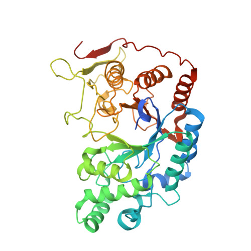
    Fucoidanases (EC 3.2.1.-) catalyze the hydrolysis of glycosidic bonds between fucose residues in fucoidans. Fucoidans are a compositionally and structurally diverse class of fucose-containing sulfated polysaccharides that are primarily found in brown seaweeds. Here, the structural characterization of a novel endo-α(1,4)-fucoidanase, Mef1, from the marine bacterium Muricauda eckloniae is presented, showing sequence similarity to members of glycoside hydrolase family 107. Using carbohydrate polyacrylamide gel electrophoresis and nuclear magnetic resonance analyses, it is shown that the fucoidanase Mef1 catalyzes the cleavage of α(1,4)-linkages between fucose residues sulfated on C2 in the structure [-3)-α-L-Fucp2S-(1,4)-α-L-Fucp2S-(1-] n in fucoidan from Fucus evanescens. Kinetic analysis of Mef1 activity by Fourier transform infrared spectroscopy revealed that the specific Mef1 fucoidanase activity (U f ) on F. evanescens fucoidan was 0.1 × 10 -3  U f  µM -1 . By crystal structure determination of Mef1 at 1.8 Å resolution, a single-domain organization comprising a (β/α) 8 -barrel domain was determined. The active site was in an extended, positively charged groove that is likely to be designed to accommodate the binding of the negatively charged, sulfated fucoidan substrate. The active site of Mef1 comprises the amino acids His270 and Asp187, providing acid/base and nucleophile groups, respectively, for the hydrolysis of glycosidic bonds in the fucoidan backbone. Electron densities were identified for two possible Ca 2+ ions in the enzyme, one of which is partially exposed to the active-site groove, while the other is very tightly coordinated. A water wire was discovered leading from the exterior of the Mef1 enzyme into the active site, passing the tightly coordinated Ca 2+ site.


  • Organizational Affiliation
    • Protein Chemistry and Enzyme Technology Section, Department of Biotechnology and Biomedicine, Technical University of Denmark, DK-2800 Kgs Lyngby, Denmark.

Macromolecule Content 

  • Total Structure Weight: 46 kDa 
  • Atom Count: 3,389 
  • Modeled Residue Count: 370 
  • Deposited Residue Count: 399 
  • Unique protein chains: 1

Macromolecules

Find similar proteins by:|  3D Structure
Entity ID: 1
MoleculeChains  Sequence LengthOrganismDetailsImage
Glycosyl hydrolase399Flagellimonas eckloniaeMutation(s): 0 
Gene Names: AAY42_01290
EC: 3.2.1.212
UniProt
Find proteins for A0A0N8WFH1 (Flagellimonas eckloniae)
Explore A0A0N8WFH1 
Go to UniProtKB:  A0A0N8WFH1
Entity Groups
Sequence Clusters30% Identity50% Identity70% Identity90% Identity95% Identity100% Identity
UniProt GroupA0A0N8WFH1
Sequence Annotations
Expand
Reference Sequence

Small Molecules

Ligands 3 Unique
IDChains Name / Formula / InChI Key2D Diagram3D Interactions
3CX
(Subject of Investigation/LOI)

Query on 3CX


Download:Ideal Coordinates CCD File
B [auth A],
D [auth A],
E [auth A]
(2S)-3-(cyclohexylamino)-2-hydroxypropane-1-sulfonic acid
C9 H19 N O4 S
INEWUCPYEUEQTN-VIFPVBQESA-N
EDO

Query on EDO


Download:Ideal Coordinates CCD File
C [auth A],
F [auth A]
1,2-ETHANEDIOL
C2 H6 O2
LYCAIKOWRPUZTN-UHFFFAOYSA-N
CA

Query on CA


Download:Ideal Coordinates CCD File
G [auth A],
H [auth A]
CALCIUM ION
Ca
BHPQYMZQTOCNFJ-UHFFFAOYSA-N

Experimental Data & Validation

Experimental Data

  • Method: X-RAY DIFFRACTION
  • Resolution: 1.80 Å
  • R-Value Free:  0.202 (Depositor), 0.203 (DCC) 
  • R-Value Work:  0.165 (Depositor), 0.166 (DCC) 
  • R-Value Observed: 0.167 (Depositor) 
Space Group: P 63
Unit Cell:
Length ( Å )Angle ( ˚ )
a = 105.416α = 90
b = 105.416β = 90
c = 78.247γ = 120
Software Package:
Software NamePurpose
PHENIXrefinement
xia2data reduction
DIALSdata scaling
PHASERphasing

Structure Validation

View Full Validation Report


Ligand Structure Quality Assessment 


Entry History 

& Funding Information

Deposition Data


Funding OrganizationLocationGrant Number
Techical University of Denmark (DTU)Denmark9082-00021B

Revision History  (Full details and data files)

  • Version 1.0: 2023-11-08
    Type: Initial release
  • Version 1.1: 2023-11-15
    Changes: Database references
  • Version 1.2: 2026-03-04
    Changes: Refinement description, Structure summary