N-terminal domain of polypyrimidine-tract binding protein is a dynamic folding platform for adaptive RNA recognition.
Damberger, F.F., Krepl, M., Arora, R., Beusch, I., Maris, C., Dorn, G., Sponer, J., Ravindranathan, S., Allain, F.H.(2024) Nucleic Acids Res 52: 10683-10704
- PubMed: 39180402 Search on PubMed
- DOI: https://doi.org/10.1093/nar/gkae713
- Primary Citation Related Structures:
8BGF - PubMed Abstract:
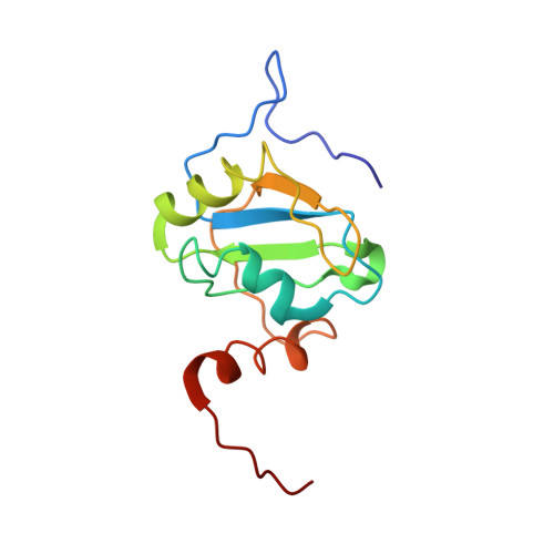
The N-terminal RNA recognition motif domain (RRM1) of polypyrimidine tract binding protein (PTB) forms an additional C-terminal helix α3, which docks to one edge of the β-sheet upon binding to a stem-loop RNA containing a UCUUU pentaloop. Importantly, α3 does not contact the RNA. The α3 helix therefore represents an allosteric means to regulate the conformation of adjacent domains in PTB upon binding structured RNAs. Here we investigate the process of dynamic adaptation by stem-loop RNA and RRM1 using NMR and MD in order to obtain mechanistic insights on how this allostery is achieved. Relaxation data and NMR structure determination of the free protein show that α3 is partially ordered and interacts with the domain transiently. Stem-loop RNA binding quenches fast time scale dynamics and α3 becomes ordered, however microsecond dynamics at the protein-RNA interface is observed. MD shows how RRM1 binding to the stem-loop RNA is coupled to the stabilization of the C-terminal helix and helps to transduce differences in RNA loop sequence into changes in α3 length and order. IRES assays of full length PTB and a mutant with altered dynamics in the α3 region show that this dynamic allostery influences PTB function in cultured HEK293T cells.
- Institute of Biochemistry, ETH Zurich, 8093 Zurich, Switzerland.
Organizational Affiliation: