The adaptability of the ion-binding site by the Ag(I)/Cu(I) periplasmic chaperone SilF.
Lithgo, R.M., Hanzevacki, M., Harris, G., Kamps, J.J.A.G., Holden, E., Gianga, T.M., Benesch, J.L.P., Jager, C.M., Croft, A.K., Hussain, R., Hobman, J.L., Orville, A.M., Quigley, A., Carr, S.B., Scott, D.J.(2023) J Biological Chem 299: 105331-105331
- PubMed: 37820867 Search on PubMedSearch on PubMed Central
- DOI: https://doi.org/10.1016/j.jbc.2023.105331
- Primary Citation Related Structures:
8BBZ, 8BWV - PubMed Abstract:
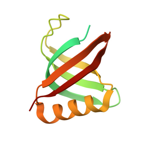
The periplasmic chaperone SilF has been identified as part of an Ag(I) detoxification system in Gram-negative bacteria. Sil proteins also bind Cu(I) but with reported weaker affinity, therefore leading to the designation of a specific detoxification system for Ag(I). Using isothermal titration calorimetry, we show that binding of both ions is not only tighter than previously thought but of very similar affinities. We investigated the structural origins of ion binding using molecular dynamics and QM/MM simulations underpinned by structural and biophysical experiments. The results of this analysis showed that the binding site adapts to accommodate either ion, with key interactions with the solvent in the case of Cu(I). The implications of this are that Gram-negative bacteria do not appear to have evolved a specific Ag(I) efflux system but take advantage of the existing Cu(I) detoxification system. Therefore, there are consequences for how we define a particular metal resistance mechanism and understand its evolution in the environment.
- School of Biosciences, Sutton Bonington Campus, University of Nottingham, Leicestershire, United Kingdom; Membrane Protein Laboratory, Diamond Light Source, Rutherford Appleton Laboratory, Didcot, Oxfordshire, United Kingdom; Diamond Light Source, Diamond House, Rutherford Appleton Laboratories, Didcot, Oxfordshire, United Kingdom; Research Complex at Harwell, Rutherford Appleton Laboratory, Didcot, Oxfordshire, United Kingdom.
Organizational Affiliation: