8BBT | pdb_00008bbt

Native Tipula oleracea Nudivirus polyhedrin - 1960


Experimental Data Snapshot

  • Method: X-RAY DIFFRACTION
  • Resolution: 1.69 Å
  • R-Value Free: 
    0.209 (Depositor), 0.207 (DCC) 
  • R-Value Work: 
    0.189 (Depositor), 0.189 (DCC) 
  • R-Value Observed: 
    0.191 (Depositor) 

Starting Model: experimental
View more details

wwPDB Validation 3D Report Full Report

Validation slider image for 8BBT

This is version 1.2 of the entry. See complete history

Literature

Atomic structure of a nudivirus occlusion body protein determined from a 70-year-old crystal sample.

Keown, J.R.Crawshaw, A.D.Trincao, J.Carrique, L.Gildea, R.J.Horrell, S.Warren, A.J.Axford, D.Owen, R.Evans, G.Bezier, A.Metcalf, P.Grimes, J.M.

(2023) Nat Commun 14: 4160-4160

  • DOI: https://doi.org/10.1038/s41467-023-39819-1
  • Primary Citation Related Structures: 
    8BBT, 8BC5, 8BCK, 8BCL

  • PubMed Abstract: 

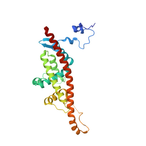
    Infectious protein crystals are an essential part of the viral lifecycle for double-stranded DNA Baculoviridae and double-stranded RNA cypoviruses. These viral protein crystals, termed occlusion bodies or polyhedra, are dense protein assemblies that form a crystalline array, encasing newly formed virions. Here, using X-ray crystallography we determine the structure of a polyhedrin from Nudiviridae. This double-stranded DNA virus family is a sister-group to the baculoviruses, whose members were thought to lack occlusion bodies. The 70-year-old sample contains a well-ordered lattice formed by a predominantly α-helical building block that assembles into a dense, highly interconnected protein crystal. The lattice is maintained by extensive hydrophobic and electrostatic interactions, disulfide bonds, and domain switching. The resulting lattice is resistant to most environmental stresses. Comparison of this structure to baculovirus or cypovirus polyhedra shows a distinct protein structure, crystal space group, and unit cell dimensions, however, all polyhedra utilise common principles of occlusion body assembly.


  • Organizational Affiliation
    • Division of Structural Biology, Wellcome Centre for Human Genetics, University of Oxford, Oxford, UK. Jeremy@strubi.ox.ac.uk.

Macromolecule Content 

  • Total Structure Weight: 27.42 kDa 
  • Atom Count: 1,964 
  • Modeled Residue Count: 230 
  • Deposited Residue Count: 241 
  • Unique protein chains: 1

Macromolecules

Find similar proteins by:|  3D Structure
Entity ID: 1
MoleculeChains  Sequence LengthOrganismDetailsImage
MOBP241Tipula oleracea nudivirusMutation(s): 0 
UniProt
Find proteins for A0A0B4VFQ3 (Tipula oleracea nudivirus)
Explore A0A0B4VFQ3 
Go to UniProtKB:  A0A0B4VFQ3
Entity Groups
Sequence Clusters30% Identity50% Identity70% Identity90% Identity95% Identity100% Identity
UniProt GroupA0A0B4VFQ3
Sequence Annotations
Expand
Reference Sequence

Small Molecules

Ligands 1 Unique
IDChains Name / Formula / InChI Key2D Diagram3D Interactions
CA

Query on CA


Download:Ideal Coordinates CCD File
B [auth A]CALCIUM ION
Ca
BHPQYMZQTOCNFJ-UHFFFAOYSA-N

Experimental Data & Validation

Experimental Data

  • Method: X-RAY DIFFRACTION
  • Resolution: 1.69 Å
  • R-Value Free:  0.209 (Depositor), 0.207 (DCC) 
  • R-Value Work:  0.189 (Depositor), 0.189 (DCC) 
  • R-Value Observed: 0.191 (Depositor) 
Space Group: P 32 2 1
Unit Cell:
Length ( Å )Angle ( ˚ )
a = 53.389α = 90
b = 53.389β = 90
c = 105.578γ = 120
Software Package:
Software NamePurpose
PHENIXrefinement
Cootmodel building
xia2.multiplexdata reduction
DIALSdata reduction
xia2.multiplexdata scaling
DIALSdata scaling
CRANKphasing

Structure Validation

View Full Validation Report


Entry History 

& Funding Information

Deposition Data


Funding OrganizationLocationGrant Number
Wellcome TrustUnited Kingdom200835/Z/16/Z

Revision History  (Full details and data files)

  • Version 1.0: 2023-07-12
    Type: Initial release
  • Version 1.1: 2024-01-24
    Changes: Data collection, Database references
  • Version 1.2: 2024-11-20
    Changes: Structure summary