8BAC | pdb_00008bac

Crystal structure of human heparanase in complex with competitive inhibitor GD05


Experimental Data Snapshot

  • Method: X-RAY DIFFRACTION
  • Resolution: 2.05 Å
  • R-Value Free: 
    0.267 (Depositor), 0.272 (DCC) 
  • R-Value Work: 
    0.224 (Depositor), 0.230 (DCC) 

Starting Model: experimental
View more details

wwPDB Validation 3D Report Full Report

Validation slider image for 8BAC

Ligand Structure Quality Assessment 


This is version 1.2 of the entry. See complete history

Literature

Synthesis of Uronic Acid 1-Azasugars as Putative Inhibitors of alpha-Iduronidase, beta-Glucuronidase and Heparanase.

Doherty, G.G.Ler, G.J.M.Wimmer, N.Bernhardt, P.V.Ashmus, R.A.Vocadlo, D.J.Armstrong, Z.Davies, G.J.Maccarana, M.Li, J.P.Kayal, Y.Ferro, V.

(2023) Chembiochem 24: e202200619-e202200619

  • DOI: https://doi.org/10.1002/cbic.202200619
  • Primary Citation Related Structures: 
    8BAC

  • PubMed Abstract: 

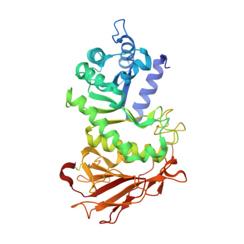
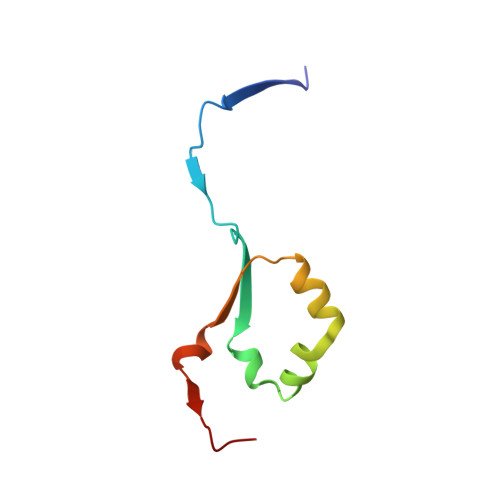
    1-Azasugar analogues of l-iduronic acid (l-IdoA) and d-glucuronic acid (d-GlcA) and their corresponding enantiomers have been synthesized as potential pharmacological chaperones for mucopolysaccharidosis I (MPS I), a lysosomal storage disease caused by mutations in the gene encoding α-iduronidase (IDUA). The compounds were efficiently synthesized in nine or ten steps from d- or l-arabinose, and the structures were confirmed by X-ray crystallographic analysis of key intermediates. All compounds were inactive against IDUA, although l-IdoA-configured 8 moderately inhibited β-glucuronidase (β-GLU). The d-GlcA-configured 9 was a potent inhibitor of β-GLU and a moderate inhibitor of the endo-β-glucuronidase heparanase. Co-crystallization of 9 with heparanase revealed that the endocyclic nitrogen of 9 forms close interactions with both the catalytic acid and catalytic nucleophile.


  • Organizational Affiliation
    • School of Chemistry & Molecular Biosciences, The University of Queensland, Brisbane, Queensland, 4072, Australia.

Macromolecule Content 

  • Total Structure Weight: 52.67 kDa 
  • Atom Count: 3,764 
  • Modeled Residue Count: 459 
  • Deposited Residue Count: 463 
  • Unique protein chains: 2

Macromolecules

Find similar proteins by:|  3D Structure
Entity ID: 1
MoleculeChains  Sequence LengthOrganismDetailsImage
Heparanase 50 kDa subunitA [auth AAA]389Homo sapiensMutation(s): 1 
Gene Names: HPSEHEPHPAHPA1HPR1HPSE1HSE1
EC: 3.2.1.166
UniProt & NIH Common Fund Data Resources
Find proteins for Q9Y251 (Homo sapiens)
Explore Q9Y251 
Go to UniProtKB:  Q9Y251
PHAROS:  Q9Y251
GTEx:  ENSG00000173083 
Entity Groups
Sequence Clusters30% Identity50% Identity70% Identity90% Identity95% Identity100% Identity
UniProt GroupQ9Y251
Glycosylation
Glycosylation Sites: 2Go to GlyGen: Q9Y251-1
Sequence Annotations
Expand
Reference Sequence
Find similar proteins by:|  3D Structure
Entity ID: 2
MoleculeChains  Sequence LengthOrganismDetailsImage
Heparanase 8 kDa subunitB [auth BBB]74Homo sapiensMutation(s): 0 
Gene Names: HPSEHEPHPAHPA1HPR1HPSE1HSE1
EC: 3.2.1.166
UniProt & NIH Common Fund Data Resources
Find proteins for Q9Y251 (Homo sapiens)
Explore Q9Y251 
Go to UniProtKB:  Q9Y251
PHAROS:  Q9Y251
GTEx:  ENSG00000173083 
Entity Groups
Sequence Clusters30% Identity50% Identity70% Identity90% Identity95% Identity100% Identity
UniProt GroupQ9Y251
Sequence Annotations
Expand
Reference Sequence

Experimental Data & Validation

Experimental Data

  • Method: X-RAY DIFFRACTION
  • Resolution: 2.05 Å
  • R-Value Free:  0.267 (Depositor), 0.272 (DCC) 
  • R-Value Work:  0.224 (Depositor), 0.230 (DCC) 
Space Group: P 1 21 1
Unit Cell:
Length ( Å )Angle ( ˚ )
a = 44.838α = 90
b = 71.188β = 98.375
c = 78.628γ = 90
Software Package:
Software NamePurpose
REFMACrefinement
xia2data reduction
xia2data scaling
PHASERphasing

Structure Validation

View Full Validation Report


Ligand Structure Quality Assessment 


Entry History 

& Funding Information

Deposition Data


Funding OrganizationLocationGrant Number
Biotechnology and Biological Sciences Research Council (BBSRC)United KingdomKen murray professorship

Revision History  (Full details and data files)

  • Version 1.0: 2023-03-01
    Type: Initial release
  • Version 1.1: 2024-02-07
    Changes: Data collection, Refinement description
  • Version 1.2: 2024-10-16
    Changes: Structure summary