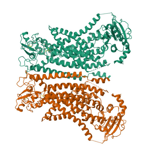
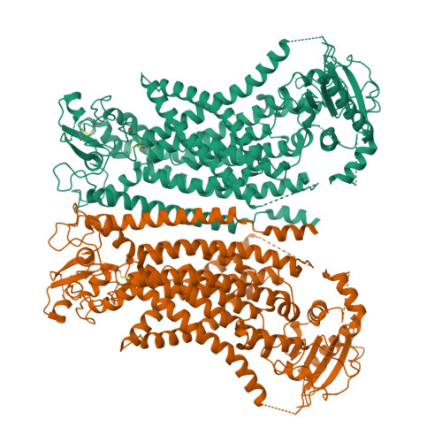
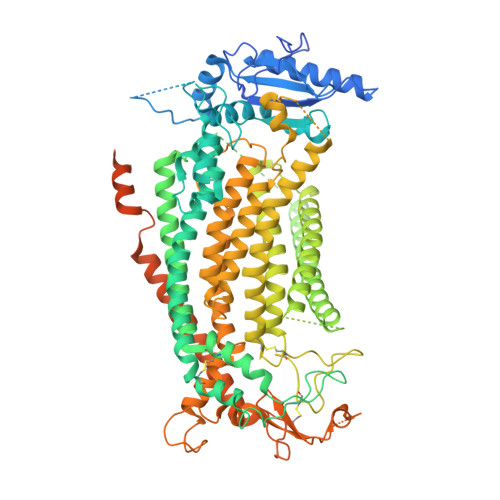
8B8Q

Structure of mTMEM16F in lipid Nanodiscs in the presence of Ca2+


Experimental Data Snapshot

  • Method: ELECTRON MICROSCOPY
  • Resolution: 2.94 Å
  • Aggregation State: PARTICLE 
  • Reconstruction Method: SINGLE PARTICLE 

Starting Model: experimental
View more details

wwPDB Validation   3D Report Full Report


This is version 1.1 of the entry. See complete history


Literature

Structural basis for the activation of the lipid scramblase TMEM16F.

Arndt, M.Alvadia, C.Straub, M.S.Clerico Mosina, V.Paulino, C.Dutzler, R.

(2022) Nat Commun 13: 6692-6692

  • DOI: https://doi.org/10.1038/s41467-022-34497-x
  • Primary Citation of Related Structures:  
    8B8G, 8B8J, 8B8K, 8B8M, 8B8Q, 8BC0, 8BC1

  • PubMed Abstract: 

    TMEM16F, a member of the conserved TMEM16 family, plays a central role in the initiation of blood coagulation and the fusion of trophoblasts. The protein mediates passive ion and lipid transport in response to an increase in intracellular Ca 2+ . However, the mechanism of how the protein facilitates both processes has remained elusive. Here we investigate the basis for TMEM16F activation. In a screen of residues lining the proposed site of conduction, we identify mutants with strongly activating phenotype. Structures of these mutants determined herein by cryo-electron microscopy show major rearrangements leading to the exposure of hydrophilic patches to the membrane, whose distortion facilitates lipid diffusion. The concomitant opening of a pore promotes ion conduction in the same protein conformation. Our work has revealed a mechanism that is distinct for this branch of the family and that will aid the development of a specific pharmacology for a promising drug target.


  • Organizational Affiliation

    Department of Biochemistry University of Zurich, Winterthurer Str. 190, CH-8057, Zurich, Switzerland.


Macromolecules
Find similar proteins by:  (by identity cutoff)  |  3D Structure
Entity ID: 1
MoleculeChains Sequence LengthOrganismDetailsImage
Anoctamin-6
A, B
975Mus musculusMutation(s): 1 
Gene Names: Ano6Tmem16f
Membrane Entity: Yes 
UniProt & NIH Common Fund Data Resources
Find proteins for Q6P9J9 (Mus musculus)
Explore Q6P9J9 
Go to UniProtKB:  Q6P9J9
IMPC:  MGI:2145890
Entity Groups  
Sequence Clusters30% Identity50% Identity70% Identity90% Identity95% Identity100% Identity
UniProt GroupQ6P9J9
Sequence Annotations
Expand
  • Reference Sequence
Experimental Data & Validation

Experimental Data

  • Method: ELECTRON MICROSCOPY
  • Resolution: 2.94 Å
  • Aggregation State: PARTICLE 
  • Reconstruction Method: SINGLE PARTICLE 
EM Software:
TaskSoftware PackageVersion
RECONSTRUCTIONcryoSPARC
MODEL REFINEMENTPHENIX

Structure Validation

View Full Validation Report


Entry History & Funding Information

Deposition Data


Funding OrganizationLocationGrant Number
European Research Council (ERC)European Union339116

Revision History  (Full details and data files)

  • Version 1.0: 2022-12-21
    Type: Initial release
  • Version 1.1: 2024-11-13
    Changes: Data collection, Refinement description, Structure summary