8B6N | pdb_00008b6n

X-ray structure of the haloalkane dehalogenase HaloTag7 circular permutated at positions 141-156 (cpHaloTagDelta)


Experimental Data Snapshot

  • Method: X-RAY DIFFRACTION
  • Resolution: 2.30 Å
  • R-Value Free: 
    0.250 (Depositor), 0.253 (DCC) 
  • R-Value Work: 
    0.215 (Depositor), 0.215 (DCC) 
  • R-Value Observed: 
    0.216 (Depositor) 

Starting Model: experimental
View more details

wwPDB Validation 3D Report Full Report

Validation slider image for 8B6N

This is version 1.3 of the entry. See complete history

Literature

Recording physiological history of cells with chemical labeling.

Huppertz, M.C.Wilhelm, J.Grenier, V.Schneider, M.W.Falt, T.Porzberg, N.Hausmann, D.Hoffmann, D.C.Hai, L.Tarnawski, M.Pino, G.Slanchev, K.Kolb, I.Acuna, C.Fenk, L.M.Baier, H.Hiblot, J.Johnsson, K.

(2024) Science 383: 890-897

  • DOI: https://doi.org/10.1126/science.adg0812
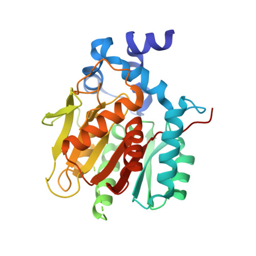
  • Primary Citation Related Structures: 
    8B6N, 8B6P

  • PubMed Abstract: 

    Recordings of the physiological history of cells provide insights into biological processes, yet obtaining such recordings is a challenge. To address this, we introduce a method to record transient cellular events for later analysis. We designed proteins that become labeled in the presence of both a specific cellular activity and a fluorescent substrate. The recording period is set by the presence of the substrate, whereas the cellular activity controls the degree of the labeling. The use of distinguishable substrates enabled the recording of successive periods of activity. We recorded protein-protein interactions, G protein-coupled receptor activation, and increases in intracellular calcium. Recordings of elevated calcium levels allowed selections of cells from heterogeneous populations for transcriptomic analysis and tracking of neuronal activities in flies and zebrafish.


  • Organizational Affiliation
    • Department of Chemical Biology, Max Planck Institute for Medical Research, Jahnstrasse 29, 69120 Heidelberg, Germany.

Macromolecule Content 

  • Total Structure Weight: 32.78 kDa 
  • Atom Count: 2,189 
  • Modeled Residue Count: 272 
  • Deposited Residue Count: 294 
  • Unique protein chains: 1

Macromolecules

Find similar proteins by:|  3D Structure
Entity ID: 1
MoleculeChains  Sequence LengthOrganismDetailsImage
Haloalkane dehalogenase294Rhodococcus sp. (in: high G+C Gram-positive bacteria)Mutation(s): 0 
Gene Names: dhaA
EC: 3.8.1.5
UniProt
Find proteins for P0A3G3 (Rhodococcus sp)
Explore P0A3G3 
Go to UniProtKB:  P0A3G3
Entity Groups
Sequence Clusters30% Identity50% Identity70% Identity90% Identity95% Identity100% Identity
UniProt GroupP0A3G3
Sequence Annotations
Expand
Reference Sequence

Small Molecules

Ligands 1 Unique
IDChains Name / Formula / InChI Key2D Diagram3D Interactions
NHE

Query on NHE


Download:Ideal Coordinates CCD File
B [auth A]2-[N-CYCLOHEXYLAMINO]ETHANE SULFONIC ACID
C8 H17 N O3 S
MKWKNSIESPFAQN-UHFFFAOYSA-N

Experimental Data & Validation

Experimental Data

  • Method: X-RAY DIFFRACTION
  • Resolution: 2.30 Å
  • R-Value Free:  0.250 (Depositor), 0.253 (DCC) 
  • R-Value Work:  0.215 (Depositor), 0.215 (DCC) 
  • R-Value Observed: 0.216 (Depositor) 
Space Group: C 2 2 21
Unit Cell:
Length ( Å )Angle ( ˚ )
a = 45.85α = 90
b = 79.32β = 90
c = 154.18γ = 90
Software Package:
Software NamePurpose
PHENIXrefinement
XDSdata reduction
XSCALEdata scaling
PHASERphasing

Structure Validation

View Full Validation Report


Entry History 

& Funding Information

Deposition Data


Funding OrganizationLocationGrant Number
Max Planck SocietyGermany--

Revision History  (Full details and data files)

  • Version 1.0: 2023-10-11
    Type: Initial release
  • Version 1.1: 2024-02-14
    Changes: Database references
  • Version 1.2: 2024-03-06
    Changes: Database references
  • Version 1.3: 2026-03-04
    Changes: Refinement description, Structure summary