8B5B | pdb_00008b5b

Human BRD4 bromdomain 1 in complex with a H4 peptide containing acetyl lysine and ApmTri (H4K5acK8ApmTri)


Experimental Data Snapshot

  • Method: X-RAY DIFFRACTION
  • Resolution: 1.92 Å
  • R-Value Free: 
    0.246 (Depositor), 0.245 (DCC) 
  • R-Value Work: 
    0.197 (Depositor), 0.198 (DCC) 
  • R-Value Observed: 
    0.199 (Depositor) 

Starting Model: experimental
View more details

wwPDB Validation 3D Report Full Report

Validation slider image for 8B5B

This is version 2.2 of the entry. See complete history

Literature

Synthesis, Biochemical Characterization, and Genetic Encoding of a 1,2,4-Triazole Amino Acid as an Acetyllysine Mimic for Bromodomains of the BET Family.

Kirchgassner, S.Braun, M.B.Bartlick, N.Koc, C.Reinkemeier, C.D.Lemke, E.A.Stehle, T.Schwarzer, D.

(2023) Angew Chem Int Ed Engl 62: e202215460-e202215460

  • DOI: https://doi.org/10.1002/anie.202215460
  • Primary Citation Related Structures: 
    8B5A, 8B5B, 8B5C

  • PubMed Abstract: 

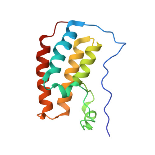
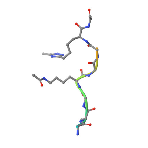
    Lysine acetylation is a charge-neutralizing post-translational modification of proteins bound by bromodomains (Brds). A 1,2,4-triazole amino acid (ApmTri) was established as acetyllysine (Kac) mimic recruiting Brds of the BET family in contrast to glutamine commonly used for simulating this modification. Optimization of triazole substituents and side chain spacing allowed BET Brd recruitment to ApmTri-containing peptides with affinities similar to native substrates. Crystal structures of ApmTri-containing peptides in complex with two BET Brds revealed the binding mode which mirrored that of Kac ligands. ApmTri was genetically encoded and recombinant ApmTri-containing proteins co-enriched BRD3(2) from cellular lysates. This interaction was blocked by BET inhibitor JQ1. With genetically encoded ApmTri, biochemistry is now provided with a stable Kac mimic reflecting charge neutralization and Brd recruitment, allowing new investigations into BET proteins in vitro and in vivo.


  • Organizational Affiliation
    • Interfakultäres Institut für Biochemie, Universität Tübingen, Auf der Morgenstelle 34, 72076, Tübingen, Germany.

Macromolecule Content 

  • Total Structure Weight: 48.46 kDa 
  • Atom Count: 3,458 
  • Modeled Residue Count: 398 
  • Deposited Residue Count: 413 
  • Unique protein chains: 2

Macromolecules

Find similar proteins by:|  3D Structure
Entity ID: 1
MoleculeChains  Sequence LengthOrganismDetailsImage
Bromodomain-containing protein 4
A, B, C
129Homo sapiensMutation(s): 0 
Gene Names: BRD4HUNK1
UniProt & NIH Common Fund Data Resources
Find proteins for O60885 (Homo sapiens)
Explore O60885 
Go to UniProtKB:  O60885
PHAROS:  O60885
GTEx:  ENSG00000141867 
Entity Groups
Sequence Clusters30% Identity50% Identity70% Identity90% Identity95% Identity100% Identity
UniProt GroupO60885
Sequence Annotations
Expand
Reference Sequence
Find similar proteins by:  Sequence   |   3D Structure  
Entity ID: 2
MoleculeChains  Sequence LengthOrganismDetailsImage
H4K5acK8ApmTri
D, E
13Homo sapiensMutation(s): 0 
UniProt & NIH Common Fund Data Resources
Find proteins for P62805 (Homo sapiens)
Explore P62805 
Go to UniProtKB:  P62805
Entity Groups
Sequence Clusters30% Identity50% Identity70% Identity90% Identity95% Identity100% Identity
UniProt GroupP62805
Sequence Annotations
Expand
Reference Sequence

Small Molecules

Ligands 1 Unique
IDChains Name / Formula / InChI Key2D Diagram3D Interactions
PEG

Query on PEG


Download:Ideal Coordinates CCD File
F [auth C]DI(HYDROXYETHYL)ETHER
C4 H10 O3
MTHSVFCYNBDYFN-UHFFFAOYSA-N
Modified Residues  1 Unique
IDChains TypeFormula2D DiagramParent
ALY
Query on ALY
D, E
L-PEPTIDE LINKINGC8 H16 N2 O3LYS

Experimental Data & Validation

Experimental Data

  • Method: X-RAY DIFFRACTION
  • Resolution: 1.92 Å
  • R-Value Free:  0.246 (Depositor), 0.245 (DCC) 
  • R-Value Work:  0.197 (Depositor), 0.198 (DCC) 
  • R-Value Observed: 0.199 (Depositor) 
Space Group: P 21 21 21
Unit Cell:
Length ( Å )Angle ( ˚ )
a = 39.508α = 90
b = 51.78β = 90
c = 170.739γ = 90
Software Package:
Software NamePurpose
PHENIXrefinement
XDSdata reduction
XDSdata scaling
PHASERphasing

Structure Validation

View Full Validation Report


Entry History 

& Funding Information

Deposition Data


Funding OrganizationLocationGrant Number
German Research Foundation (DFG)GermanySCHW 1163/9-1

Revision History  (Full details and data files)

  • Version 1.0: 2023-01-11
    Type: Initial release
  • Version 1.1: 2023-02-15
    Changes: Database references
  • Version 1.2: 2023-03-15
    Changes: Database references
  • Version 1.3: 2023-03-22
    Changes: Database references
  • Version 2.0: 2023-05-24
    Changes: Non-polymer description
  • Version 2.1: 2024-02-07
    Changes: Data collection, Derived calculations, Refinement description
  • Version 2.2: 2024-10-23
    Changes: Structure summary