8B18 | pdb_00008b18

DtpB-Nb132-AF


Experimental Data Snapshot

  • Method: X-RAY DIFFRACTION
  • Resolution: 2.30 Å
  • R-Value Free: 
    0.254 (Depositor), 0.257 (DCC) 
  • R-Value Work: 
    0.227 (Depositor) 
  • R-Value Observed: 
    0.228 (Depositor) 

Starting Model: experimental
View more details

wwPDB Validation 3D Report Full Report

Validation slider image for 8B18

Ligand Structure Quality Assessment 


This is version 1.2 of the entry. See complete history

Literature

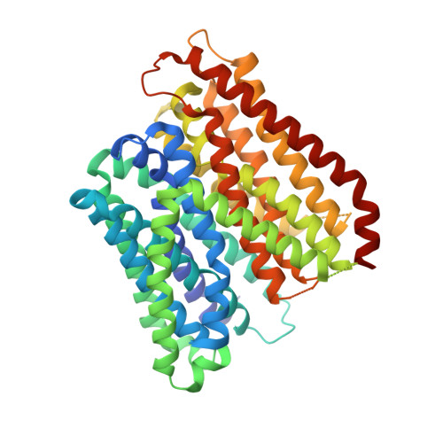
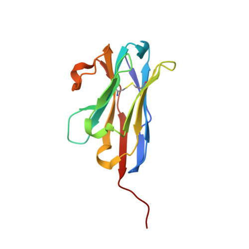
Plasticity of the binding pocket in peptide transporters underpins promiscuous substrate recognition.

Kotov, V.Killer, M.Jungnickel, K.E.J.Lei, J.Finocchio, G.Steinke, J.Bartels, K.Strauss, J.Dupeux, F.Humm, A.S.Cornaciu, I.Marquez, J.A.Pardon, E.Steyaert, J.Low, C.

(2023) Cell Rep 42: 112831-112831

  • DOI: https://doi.org/10.1016/j.celrep.2023.112831
  • Primary Citation Related Structures: 
    8B17, 8B18, 8B19, 8B1A, 8B1B, 8B1C, 8B1D, 8B1E, 8B1F, 8B1G, 8B1H, 8B1I, 8B1J, 8B1K

  • PubMed Abstract: 

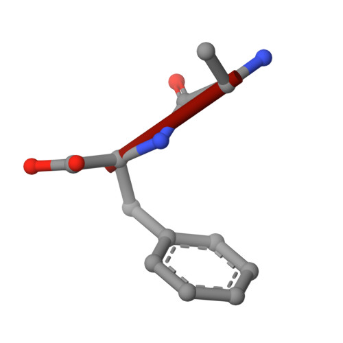
    Proton-dependent oligopeptide transporters (POTs) are promiscuous transporters of the major facilitator superfamily that constitute the main route of entry for a wide range of dietary peptides and orally administrated peptidomimetic drugs. Given their clinical and pathophysiological relevance, several POT homologs have been studied extensively at the structural and molecular level. However, the molecular basis of recognition and transport of diverse peptide substrates has remained elusive. We present 14 X-ray structures of the bacterial POT DtpB in complex with chemically diverse di- and tripeptides, providing novel insights into the plasticity of the conserved central binding cavity. We analyzed binding affinities for more than 80 peptides and monitored uptake by a fluorescence-based transport assay. To probe whether all 8400 natural di- and tripeptides can bind to DtpB, we employed state-of-the-art molecular docking and machine learning and conclude that peptides with compact hydrophobic residues are the best DtpB binders.


  • Organizational Affiliation
    • Center for Structural Systems Biology (CSSB), Notkestraße 85, 22607 Hamburg, Germany; European Molecular Biology Laboratory (EMBL) Hamburg, Notkestraße 85, 22607 Hamburg, Germany.

Macromolecule Content 

  • Total Structure Weight: 70.37 kDa 
  • Atom Count: 4,718 
  • Modeled Residue Count: 580 
  • Deposited Residue Count: 618 
  • Unique protein chains: 3

Macromolecules

Find similar proteins by:|  3D Structure
Entity ID: 1
MoleculeChains  Sequence LengthOrganismDetailsImage
Dipeptide and tripeptide permease B489Escherichia coliMutation(s): 0 
Gene Names: dtpByhiPb3496JW3463
Membrane Entity: Yes 
UniProt
Find proteins for P36837 (Escherichia coli (strain K12))
Explore P36837 
Go to UniProtKB:  P36837
Entity Groups
Sequence Clusters30% Identity50% Identity70% Identity90% Identity95% Identity100% Identity
UniProt GroupP36837
Sequence Annotations
Expand
Reference Sequence
Find similar proteins by:|  3D Structure
Entity ID: 2
MoleculeChains  Sequence LengthOrganismDetailsImage
Nanobody 132127Lama glamaMutation(s): 0 
Find similar proteins by:  Sequence   |   3D Structure  
Entity ID: 3
MoleculeChains  Sequence LengthOrganismDetailsImage
ALA-PHE2Escherichia coliMutation(s): 0 

Small Molecules

Ligands 8 Unique
IDChains Name / Formula / InChI Key2D Diagram3D Interactions
LMT
(Subject of Investigation/LOI)

Query on LMT


Download:Ideal Coordinates CCD File
D [auth A]DODECYL-BETA-D-MALTOSIDE
C24 H46 O11
NLEBIOOXCVAHBD-QKMCSOCLSA-N
1PE
(Subject of Investigation/LOI)

Query on 1PE


Download:Ideal Coordinates CCD File
G [auth A],
N [auth A]
PENTAETHYLENE GLYCOL
C10 H22 O6
JLFNLZLINWHATN-UHFFFAOYSA-N
PG4
(Subject of Investigation/LOI)

Query on PG4


Download:Ideal Coordinates CCD File
F [auth A],
H [auth A],
I [auth A],
J [auth A],
K [auth A]
TETRAETHYLENE GLYCOL
C8 H18 O5
UWHCKJMYHZGTIT-UHFFFAOYSA-N
D12
(Subject of Investigation/LOI)

Query on D12


Download:Ideal Coordinates CCD File
L [auth A]DODECANE
C12 H26
SNRUBQQJIBEYMU-UHFFFAOYSA-N
D10
(Subject of Investigation/LOI)

Query on D10


Download:Ideal Coordinates CCD File
O [auth A]DECANE
C10 H22
DIOQZVSQGTUSAI-UHFFFAOYSA-N
OCT
(Subject of Investigation/LOI)

Query on OCT


Download:Ideal Coordinates CCD File
E [auth A]N-OCTANE
C8 H18
TVMXDCGIABBOFY-UHFFFAOYSA-N
GOL
(Subject of Investigation/LOI)

Query on GOL


Download:Ideal Coordinates CCD File
M [auth A],
P [auth A]
GLYCEROL
C3 H8 O3
PEDCQBHIVMGVHV-UHFFFAOYSA-N
HEX
(Subject of Investigation/LOI)

Query on HEX


Download:Ideal Coordinates CCD File
Q [auth A],
R [auth A]
HEXANE
C6 H14
VLKZOEOYAKHREP-UHFFFAOYSA-N

Experimental Data & Validation

Experimental Data

  • Method: X-RAY DIFFRACTION
  • Resolution: 2.30 Å
  • R-Value Free:  0.254 (Depositor), 0.257 (DCC) 
  • R-Value Work:  0.227 (Depositor) 
  • R-Value Observed: 0.228 (Depositor) 
Space Group: P 2 21 21
Unit Cell:
Length ( Å )Angle ( ˚ )
a = 54.415α = 90
b = 125.074β = 90
c = 168.713γ = 90
Software Package:
Software NamePurpose
PHENIXrefinement
XDSdata reduction

Structure Validation

View Full Validation Report


Ligand Structure Quality Assessment 


Entry History 

& Funding Information

Deposition Data


Funding OrganizationLocationGrant Number
Not funded--

Revision History  (Full details and data files)

  • Version 1.0: 2023-08-09
    Type: Initial release
  • Version 1.1: 2024-11-20
    Changes: Data collection, Structure summary
  • Version 1.2: 2026-03-04
    Changes: Refinement description