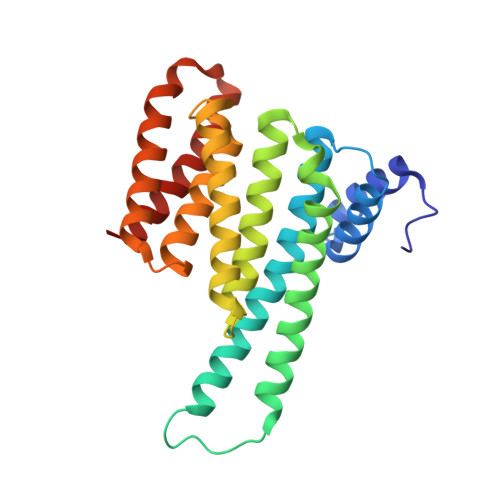
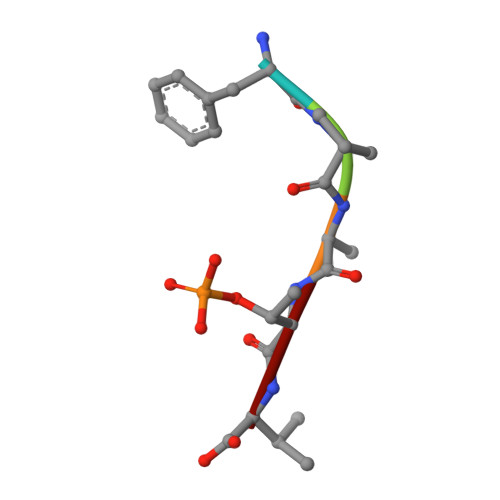
8AUS

Small molecular stabilizer for ERalpha and 14-3-3 (1080297)


Experimental Data Snapshot

  • Method: X-RAY DIFFRACTION
  • Resolution: 1.40 Å
  • R-Value Free: 0.216 
  • R-Value Work: 0.194 
  • R-Value Observed: 0.195 

wwPDB Validation   3D Report Full Report


Ligand Structure Quality Assessment 


This is version 1.2 of the entry. See complete history


Literature

Structure-Based Optimization of Covalent, Small-Molecule Stabilizers of the 14-3-3 sigma /ER alpha Protein-Protein Interaction from Nonselective Fragments.

Konstantinidou, M.Visser, E.J.Vandenboorn, E.Chen, S.Jaishankar, P.Overmans, M.Dutta, S.Neitz, R.J.Renslo, A.R.Ottmann, C.Brunsveld, L.Arkin, M.R.

(2023) J Am Chem Soc 145: 20328-20343

  • DOI: https://doi.org/10.1021/jacs.3c05161
  • Primary Citation of Related Structures:  
    8AI0, 8ALR, 8ALT, 8ALV, 8ALW, 8AM7, 8ANF, 8AOY, 8APS, 8AQ1, 8AQC, 8AQE, 8AQZ, 8AR4, 8AR5, 8ARG, 8ARO, 8ARQ, 8ARR, 8ARW, 8ARX, 8ARY, 8ARZ, 8AS1, 8AT9, 8ATP, 8ATR, 8ATS, 8AU2, 8AUS, 8AUY, 8AV3, 8AV4, 8AV7, 8AV8, 8AWG, 8AXE, 8AXU, 8AZE, 8B39

  • PubMed Abstract: 

    The stabilization of protein-protein interactions (PPIs) has emerged as a promising strategy in chemical biology and drug discovery. The identification of suitable starting points for stabilizing native PPIs and their subsequent elaboration into selective and potent molecular glues lacks structure-guided optimization strategies. We have previously identified a disulfide fragment that stabilized the hub protein 14-3-3σ bound to several of its clients, including ERα and C-RAF. Here, we show the structure-based optimization of the nonselective fragment toward selective and highly potent small-molecule stabilizers of the 14-3-3σ/ERα complex. The more elaborated molecular glues, for example, show no stabilization of 14-3-3σ/C-RAF up to 150 μM compound. Orthogonal biophysical assays, including mass spectrometry and fluorescence anisotropy, were used to establish structure-activity relationships. The binding modes of 37 compounds were elucidated with X-ray crystallography, which further assisted the concomitant structure-guided optimization. By targeting specific amino acids in the 14-3-3σ/ERα interface and locking the conformation with a spirocycle, the optimized covalent stabilizer 181 achieved potency, cooperativity, and selectivity similar to the natural product Fusicoccin-A. This case study showcases the value of addressing the structure, kinetics, and cooperativity for molecular glue development.


  • Organizational Affiliation

    Department of Pharmaceutical Chemistry and Small Molecule Discovery Center (SMDC), University of California, San Francisco, California 94143, United States.


Macromolecules
Find similar proteins by:  (by identity cutoff)  |  3D Structure
Entity ID: 1
MoleculeChains Sequence LengthOrganismDetailsImage
14-3-3 protein sigma236Homo sapiensMutation(s): 0 
Gene Names: SFNHME1
UniProt & NIH Common Fund Data Resources
Find proteins for P31947 (Homo sapiens)
Go to UniProtKB:  P31947
PHAROS:  P31947
GTEx:  ENSG00000175793 
Sequence Annotations
Expand
  • Reference Sequence

Find similar proteins by:  Sequence   |   3D Structure  

Entity ID: 2
MoleculeChains Sequence LengthOrganismDetailsImage
Estrogen receptor5Homo sapiensMutation(s): 0 
UniProt & NIH Common Fund Data Resources
Find proteins for P03372 (Homo sapiens)
Go to UniProtKB:  P03372
PHAROS:  P03372
GTEx:  ENSG00000091831 
Sequence Annotations
Expand
  • Reference Sequence
Small Molecules
Ligands 2 Unique
IDChains Name / Formula / InChI Key2D Diagram3D Interactions
O3F (Subject of Investigation/LOI)
Query on O3F

Download Ideal Coordinates CCD File 
E [auth A]2-chloranyl-~{N}-[[7-[4-[(4-chlorophenyl)amino]oxan-4-yl]carbonyl-7-azaspiro[3.5]nonan-2-yl]methyl]ethanamide
C23 H31 Cl2 N3 O3
FBJDOMAUJAZZCZ-UHFFFAOYSA-N
MG
Query on MG

Download Ideal Coordinates CCD File 
C [auth A],
D [auth A]
MAGNESIUM ION
Mg
JLVVSXFLKOJNIY-UHFFFAOYSA-N
Modified Residues  1 Unique
IDChains TypeFormula2D DiagramParent
TPO
Query on TPO
B
L-PEPTIDE LINKINGC4 H10 N O6 PTHR
Experimental Data & Validation

Experimental Data

  • Method: X-RAY DIFFRACTION
  • Resolution: 1.40 Å
  • R-Value Free: 0.216 
  • R-Value Work: 0.194 
  • R-Value Observed: 0.195 
  • Space Group: C 2 2 21
Unit Cell:
Length ( Å )Angle ( ˚ )
a = 81.879α = 90
b = 112.428β = 90
c = 62.485γ = 90
Software Package:
Software NamePurpose
REFMACrefinement
PDB_EXTRACTdata extraction
DIALSdata reduction
Aimlessdata scaling
MOLREPphasing

Structure Validation

View Full Validation Report


Ligand Structure Quality Assessment 


Entry History & Funding Information

Deposition Data


Funding OrganizationLocationGrant Number
Netherlands Organisation for Scientific Research (NWO)Netherlands--

Revision History  (Full details and data files)

  • Version 1.0: 2023-09-20
    Type: Initial release
  • Version 1.1: 2023-10-04
    Changes: Database references
  • Version 1.2: 2024-10-23
    Changes: Structure summary