8AU4 | pdb_00008au4

Structural insights reveal a heterotetramer between oncogenic K-Ras4BG12V and Rgl2, a RalA/B activator


Experimental Data Snapshot

  • Method: SOLUTION NMR
  • Conformers Calculated: 100 
  • Conformers Submitted: 20 
  • Selection Criteria: target function 

wwPDB Validation 3D Report Full Report

Validation slider image for 8AU4

This is version 1.1 of the entry. See complete history

Literature

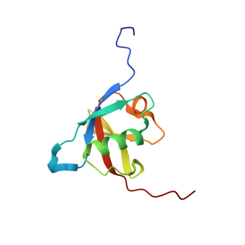
Structural insights into the complex of oncogenic KRas4B G12V and Rgl2, a RalA/B activator.

Tariq, M.Ikeya, T.Togashi, N.Fairall, L.Kamei, S.Mayooramurugan, S.Abbott, L.R.Hasan, A.Bueno-Alejo, C.Sukegawa, S.Romartinez-Alonso, B.Muro Campillo, M.A.Hudson, A.J.Ito, Y.Schwabe, J.W.Dominguez, C.Tanaka, K.

(2024) Life Sci Alliance 7

  • DOI: https://doi.org/10.26508/lsa.202302080
  • Primary Citation Related Structures: 
    8AU4, 8B69

  • PubMed Abstract: 

    About a quarter of total human cancers carry mutations in Ras isoforms. Accumulating evidence suggests that small GTPases, RalA, and RalB, and their activators, Ral guanine nucleotide exchange factors (RalGEFs), play an essential role in oncogenic Ras-induced signalling. We studied the interaction between human KRas4B and the Ras association (RA) domain of Rgl2 (Rgl2 RA ), one of the RA-containing RalGEFs. We show that the G12V oncogenic KRas4B mutation changes the interaction kinetics with Rgl2 RA The crystal structure of the KRas4B G12V : Rgl2 RA complex shows a 2:2 heterotetramer where the switch I and switch II regions of each KRas G12V interact with both Rgl2 RA molecules. This structural arrangement is highly similar to the HRas E31K :RALGDS RA crystal structure and is distinct from the well-characterised Ras:Raf complex. Interestingly, the G12V mutation was found at the dimer interface of KRas4B G12V with its partner. Our study reveals a potentially distinct mode of Ras:effector complex formation by RalGEFs and offers a possible mechanistic explanation for how the oncogenic KRas4B G12V hyperactivates the RalA/B pathway.


  • Organizational Affiliation
    • https://ror.org/04h699437 Department of Molecular and Cell Biology, University of Leicester, Leicester, UK.

Macromolecule Content 

  • Total Structure Weight: 10.97 kDa 
  • Atom Count: 766 
  • Modeled Residue Count: 100 
  • Deposited Residue Count: 100 
  • Unique protein chains: 1

Macromolecules

Find similar proteins by:|  3D Structure
Entity ID: 1
MoleculeChains  Sequence LengthOrganismDetailsImage
Ral guanine nucleotide dissociation stimulator-like 2100Homo sapiensMutation(s): 0 
Gene Names: RGL2RAB2L
UniProt & NIH Common Fund Data Resources
Find proteins for O15211 (Homo sapiens)
Explore O15211 
Go to UniProtKB:  O15211
PHAROS:  O15211
GTEx:  ENSG00000237441 
Entity Groups
Sequence Clusters30% Identity50% Identity70% Identity90% Identity95% Identity100% Identity
UniProt GroupO15211
Sequence Annotations
Expand
Reference Sequence

Experimental Data & Validation

Experimental Data

  • Method: SOLUTION NMR
  • Conformers Calculated: 100 
  • Conformers Submitted: 20 
  • Selection Criteria: target function 

Structure Validation

View Full Validation Report


Entry History 

& Funding Information

Deposition Data


Funding OrganizationLocationGrant Number
Japan Science and TechnologyJapanJPMJCR13M3, JPMJCR21E5
Japan Society for the Promotion of Science (JSPS)JapanJP15K06979, JP15K06979, JP15H01645, JP16H00847, JP17H05887, JP19H05773, JP19H05773, JP19H05773, JP19H05773, JP19H05773
Other privateShimazu foundation
Other privatethe Precise Measurement Technology Promotion Foundation

Revision History  (Full details and data files)

  • Version 1.0: 2023-08-23
    Type: Initial release
  • Version 1.1: 2023-10-25
    Changes: Database references