8ARP | pdb_00008arp

Crystal structure of DEAD-box protein Dbp2 in complex with ADP


Experimental Data Snapshot

  • Method: X-RAY DIFFRACTION
  • Resolution: 3.05 Å
  • R-Value Free: 
    0.270 (Depositor), 0.273 (DCC) 
  • R-Value Work: 
    0.223 (Depositor), 0.227 (DCC) 
  • R-Value Observed: 
    0.654 (Depositor) 

Starting Model: experimental
View more details

wwPDB Validation 3D Report Full Report

Validation slider image for 8ARP

Ligand Structure Quality Assessment 


This is version 1.3 of the entry. See complete history

Literature

Nonstructural N- and C-tails of Dbp2 confer the protein full helicase activities.

Song, Q.X.Liu, N.N.Liu, Z.X.Zhang, Y.Z.Rety, S.Hou, X.M.Xi, X.G.

(2023) J Biological Chem 299: 104592-104592

  • DOI: https://doi.org/10.1016/j.jbc.2023.104592
  • Primary Citation Related Structures: 
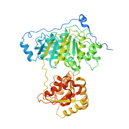
    8ARK, 8ARP

  • PubMed Abstract: 

    Human DDX5 and its yeast ortholog Dbp2 are ATP-dependent RNA helicases that play a key role in normal cell processes, cancer development, and viral infection. The crystal structure of the RecA1-like domain of DDX5 is available but the global structure of DDX5/Dbp2 subfamily proteins remains to be elucidated. Here, we report the first X-ray crystal structures of the Dbp2 helicase core alone and in complex with ADP at 3.22 Å and 3.05 Å resolutions, respectively. The structures of the ADP-bound post-hydrolysis state and apo-state demonstrate the conformational changes that occur when the nucleotides are released. Our results showed that the helicase core of Dbp2 shifted between open and closed conformation in solution but the unwinding activity was hindered when the helicase core was restricted to a single conformation. A small-angle X-ray scattering experiment showed that the disordered amino (N) tail and carboxy (C) tails are flexible in solution. Truncation mutations confirmed that the terminal tails were critical for the nucleic acid binding, ATPase, and unwinding activities, with the C-tail being exclusively responsible for the annealing activity. Furthermore, we labeled the terminal tails to observe the conformational changes between the disordered tails and the helicase core upon binding nucleic acid substrates. Specifically, we found that the nonstructural terminal tails bind to RNA substrates and tether them to the helicase core domain, thereby conferring full helicase activities to the Dbp2 protein. This distinct structural characteristic provides new insight into the mechanism of DEAD-box RNA helicases.


  • Organizational Affiliation
    • College of Life Sciences, State Key Laboratory of Crop Stress Biology in Arid Areas, Northwest A&F University, Yangling, Shaanxi, PR China.

Macromolecule Content 

  • Total Structure Weight: 369.9 kDa 
  • Atom Count: 21,294 
  • Modeled Residue Count: 2,663 
  • Deposited Residue Count: 3,276 
  • Unique protein chains: 1

Macromolecules

Find similar proteins by:|  3D Structure
Entity ID: 1
MoleculeChains  Sequence LengthOrganismDetailsImage
ATP-dependent RNA helicase DBP2
A, B, C, D, E
A, B, C, D, E, F
546Saccharomyces cerevisiaeMutation(s): 0 
Gene Names: DBP2YNL112WN1945
EC: 3.6.4.13
UniProt
Find proteins for P24783 (Saccharomyces cerevisiae (strain ATCC 204508 / S288c))
Explore P24783 
Go to UniProtKB:  P24783
Entity Groups
Sequence Clusters30% Identity50% Identity70% Identity90% Identity95% Identity100% Identity
UniProt GroupP24783
Sequence Annotations
Expand
Reference Sequence

Small Molecules

Ligands 3 Unique
IDChains Name / Formula / InChI Key2D Diagram3D Interactions
ADP
(Subject of Investigation/LOI)

Query on ADP


Download:Ideal Coordinates CCD File
G [auth A]
J [auth B]
L [auth C]
O [auth D]
Q [auth E]
G [auth A],
J [auth B],
L [auth C],
O [auth D],
Q [auth E],
V [auth F]
ADENOSINE-5'-DIPHOSPHATE
C10 H15 N5 O10 P2
XTWYTFMLZFPYCI-KQYNXXCUSA-N
SO4

Query on SO4


Download:Ideal Coordinates CCD File
H [auth A]
K [auth B]
M [auth C]
P [auth D]
R [auth E]
H [auth A],
K [auth B],
M [auth C],
P [auth D],
R [auth E],
S [auth E],
T [auth E],
W [auth F]
SULFATE ION
O4 S
QAOWNCQODCNURD-UHFFFAOYSA-L
MG
(Subject of Investigation/LOI)

Query on MG


Download:Ideal Coordinates CCD File
I [auth A],
N [auth C],
U [auth E]
MAGNESIUM ION
Mg
JLVVSXFLKOJNIY-UHFFFAOYSA-N

Experimental Data & Validation

Experimental Data

  • Method: X-RAY DIFFRACTION
  • Resolution: 3.05 Å
  • R-Value Free:  0.270 (Depositor), 0.273 (DCC) 
  • R-Value Work:  0.223 (Depositor), 0.227 (DCC) 
  • R-Value Observed: 0.654 (Depositor) 
Space Group: P 21 21 21
Unit Cell:
Length ( Å )Angle ( ˚ )
a = 145.939α = 90
b = 168.843β = 90
c = 168.87γ = 90
Software Package:
Software NamePurpose
PHENIXrefinement
PHENIXrefinement
autoPROCdata reduction
Aimlessdata scaling
PHASERphasing

Structure Validation

View Full Validation Report


Ligand Structure Quality Assessment 


Entry History 

& Funding Information

Deposition Data


Funding OrganizationLocationGrant Number
Not funded--

Revision History  (Full details and data files)

  • Version 1.0: 2023-03-22
    Type: Initial release
  • Version 1.1: 2023-05-24
    Changes: Database references
  • Version 1.2: 2024-02-07
    Changes: Data collection, Refinement description
  • Version 1.3: 2025-10-01
    Changes: Advisory, Derived calculations, Structure summary