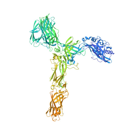
8AN2 | pdb_00008an2

S-layer protein SlaA from Sulfolobus acidocaldarius at pH 10.0


Experimental Data Snapshot

  • Method: ELECTRON MICROSCOPY
  • Resolution: 3.20 Å
  • Aggregation State: PARTICLE 
  • Reconstruction Method: SINGLE PARTICLE 

Starting Model: experimental
View more details

wwPDB Validation 3D Report Full Report

Validation slider image for 8AN2

This is version 1.2 of the entry. See complete history. 

Literature

Structure of the two-component S-layer of the archaeon Sulfolobus acidocaldarius.

Gambelli, L., McLaren, M., Conners, R., Sanders, K., Gaines, M.C., Clark, L., Gold, V.A.M., Kattnig, D., Sikora, M., Hanus, C., Isupov, M.N., Daum, B.

(2024) Elife 13

  • DOI: https://doi.org/10.7554/eLife.84617
  • Primary Citation Related Structures: 
    7ZCX, 8AN2, 8AN3, 8QOX, 8QP0

  • PubMed Abstract: 

    Surface layers (S-layers) are resilient two-dimensional protein lattices that encapsulate many bacteria and most archaea. In archaea, S-layers usually form the only structural component of the cell wall and thus act as the final frontier between the cell and its environment. Therefore, S-layers are crucial for supporting microbial life. Notwithstanding their importance, little is known about archaeal S-layers at the atomic level. Here, we combined single-particle cryo electron microscopy, cryo electron tomography, and Alphafold2 predictions to generate an atomic model of the two-component S-layer of Sulfolobus acidocaldarius . The outer component of this S-layer (SlaA) is a flexible, highly glycosylated, and stable protein. Together with the inner and membrane-bound component (SlaB), they assemble into a porous and interwoven lattice. We hypothesise that jackknife-like conformational changes in SlaA play important roles in S-layer assembly.


  • Organizational Affiliation: 
    • Living Systems Institute, University of Exeter, Exeter, United Kingdom.

Macromolecule Content 

  • Total Structure Weight: 163.78 kDa 
  • Atom Count: 8,657 
  • Modeled Residue Count: 1,040 
  • Deposited Residue Count: 1,424 
  • Unique protein chains: 1

Macromolecules

Find similar proteins by:|  3D Structure
Entity ID: 1
MoleculeChains  Sequence LengthOrganismDetailsImage
S-layer protein AA [auth AAA]1,424Sulfolobus acidocaldarius DSM 639Mutation(s): 0 
Gene Names: slaA, slp1, Saci_2355
UniProt
Find proteins for Q4J6E5 (Sulfolobus acidocaldarius (strain ATCC 33909 / DSM 639 / JCM 8929 / NBRC 15157 / NCIMB 11770))
Explore Q4J6E5 
Go to UniProtKB:  Q4J6E5
Entity Groups
Sequence Clusters30% Identity50% Identity70% Identity90% Identity95% Identity100% Identity
UniProt GroupQ4J6E5
Glycosylation
Glycosylation Sites: 19
Sequence Annotations
Expand
Reference Sequence

Oligosaccharides

Help  
Entity ID: 2
MoleculeChains Length2D Diagram GlycosylationD Interactions
alpha-D-mannopyranose-(1-6)-2-acetamido-2-deoxy-beta-D-glucopyranose-(1-4)-2-acetamido-2-deoxy-beta-D-glucopyranoseB [auth BBB]3N-Glycosylation
Glycosylation Resources
GlyTouCan: G34031ES
GlyCosmos: G34031ES
GlyGen: G34031ES
Entity ID: 3
MoleculeChains Length2D Diagram GlycosylationD Interactions
2-acetamido-2-deoxy-beta-D-glucopyranose-(1-4)-2-acetamido-2-deoxy-beta-D-glucopyranoseC [auth A],
E [auth C],
O [auth M],
Q [auth O]
2N-Glycosylation
Glycosylation Resources
GlyTouCan: G42666HT
GlyCosmos: G42666HT
GlyGen: G42666HT
Entity ID: 4
MoleculeChains Length2D Diagram GlycosylationD Interactions
beta-D-glucopyranose-(1-4)-6-deoxy-6-sulfo-beta-D-glucopyranose-(1-3)-[alpha-D-mannopyranose-(1-4)][alpha-D-mannopyranose-(1-6)]2-acetamido-2-deoxy-beta-D-glucopyranose-(1-4)-2-acetamido-2-deoxy-beta-D-glucopyranoseD [auth B]6N/AN-Glycosylation
Entity ID: 5
MoleculeChains Length2D Diagram GlycosylationD Interactions
6-deoxy-6-sulfo-beta-D-glucopyranose-(1-3)-2-acetamido-2-deoxy-beta-D-glucopyranose-(1-4)-2-acetamido-2-deoxy-beta-D-glucopyranoseF [auth D],
G [auth E],
J [auth H]
3N/AN-Glycosylation
Entity ID: 6
MoleculeChains Length2D Diagram GlycosylationD Interactions
6-deoxy-6-sulfo-beta-D-glucopyranose-(1-3)-[alpha-D-mannopyranose-(1-4)][alpha-D-mannopyranose-(1-6)]2-acetamido-2-deoxy-beta-D-glucopyranose-(1-4)-2-acetamido-2-deoxy-beta-D-glucopyranose
H [auth F],
I [auth G],
L [auth J],
M [auth K],
N [auth L],
H [auth F],
I [auth G],
L [auth J],
M [auth K],
N [auth L],
P [auth N]
5N/AN-Glycosylation
Entity ID: 7
MoleculeChains Length2D Diagram GlycosylationD Interactions
6-deoxy-6-sulfo-beta-D-glucopyranose-(1-3)-[alpha-D-mannopyranose-(1-6)]2-acetamido-2-deoxy-beta-D-glucopyranose-(1-4)-2-acetamido-2-deoxy-beta-D-glucopyranoseK [auth I]4N/AN-Glycosylation

Experimental Data & Validation

Experimental Data

  • Method: ELECTRON MICROSCOPY
  • Resolution: 3.20 Å
  • Aggregation State: PARTICLE 
  • Reconstruction Method: SINGLE PARTICLE 
EM Software:
TaskSoftware PackageVersion
MODEL REFINEMENTREFMAC

Structure Validation

View Full Validation Report


Entry History 

& Funding Information

Deposition Data


Funding OrganizationLocationGrant Number
European Research Council (ERC)European Union803894

Revision History  (Full details and data files)

  • Version 1.0: 2023-08-16
    Type: Initial release
  • Version 1.1: 2024-02-21
    Changes: Database references, Refinement description
  • Version 1.2: 2024-11-13
    Changes: Data collection, Structure summary