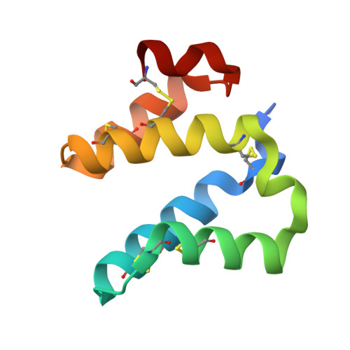
8AMY

High-resolution crystal structure of the Mu8.1 conotoxin from Conus Mucronatus


Experimental Data Snapshot

  • Method: X-RAY DIFFRACTION
  • Resolution: 1.67 Å
  • R-Value Free: 0.200 
  • R-Value Work: 0.169 
  • R-Value Observed: 0.170 

Starting Model: experimental
View more details

wwPDB Validation   3D Report Full Report


This is version 1.3 of the entry. See complete history


Literature

High-resolution crystal structure of the Mu8.1 conotoxin from Conus mucronatus.

Muller, E.Hackney, C.M.Ellgaard, L.Morth, J.P.

(2023) Acta Crystallogr F Struct Biol Commun 79: 240-246

  • DOI: https://doi.org/10.1107/S2053230X23007070
  • Primary Citation of Related Structures:  
    8AMY

  • PubMed Abstract: 

    Marine cone snails produce a wealth of peptide toxins (conotoxins) that bind their molecular targets with high selectivity and potency. Therefore, conotoxins constitute valuable biomolecular tools with a variety of biomedical purposes. The Mu8.1 conotoxin from Conus mucronatus is the founding member of the newly identified saposin-like conotoxin class of conotoxins and has been shown to target Cav2.3, a voltage-gated calcium channel. Two crystal structures have recently been determined of Mu8.1 at 2.3 and 2.1 Å resolution. Here, a high-resolution crystal structure of Mu8.1 was determined at 1.67 Å resolution in the high-symmetry space group I4 1 22. The asymmetric unit contained one molecule, with a symmetry-related molecule generating a dimer equivalent to that observed in the two previously determined structures. The high resolution allows a detailed atomic analysis of a water-filled cavity buried at the dimer interface, revealing a tightly coordinated network of waters that shield a lysine residue (Lys55) with a predicted unusually low side-chain pK a value. These findings are discussed in terms of a potential functional role of Lys55 in target interaction.


  • Organizational Affiliation

    Department of Biotechnology and Biomedicine, Technical University of Denmark, 2800 Kongens Lyngby, Denmark.


Macromolecules
Find similar proteins by:  (by identity cutoff)  |  3D Structure
Entity ID: 1
MoleculeChains Sequence LengthOrganismDetailsImage
Mu8.1 conotoxin89Conus mucronatusMutation(s): 0 
UniProt
Find proteins for P0DRC4 (Conus mucronatus)
Explore P0DRC4 
Go to UniProtKB:  P0DRC4
Entity Groups  
Sequence Clusters30% Identity50% Identity70% Identity90% Identity95% Identity100% Identity
UniProt GroupP0DRC4
Sequence Annotations
Expand
  • Reference Sequence
Small Molecules
Ligands 6 Unique
IDChains Name / Formula / InChI Key2D Diagram3D Interactions
MES
Query on MES

Download Ideal Coordinates CCD File 
B [auth A]2-(N-MORPHOLINO)-ETHANESULFONIC ACID
C6 H13 N O4 S
SXGZJKUKBWWHRA-UHFFFAOYSA-N
P4G
Query on P4G

Download Ideal Coordinates CCD File 
C [auth A]1-ETHOXY-2-(2-ETHOXYETHOXY)ETHANE
C8 H18 O3
RRQYJINTUHWNHW-UHFFFAOYSA-N
SO4
Query on SO4

Download Ideal Coordinates CCD File 
E [auth A],
F [auth A]
SULFATE ION
O4 S
QAOWNCQODCNURD-UHFFFAOYSA-L
ZN (Subject of Investigation/LOI)
Query on ZN

Download Ideal Coordinates CCD File 
G [auth A]ZINC ION
Zn
PTFCDOFLOPIGGS-UHFFFAOYSA-N
BCT
Query on BCT

Download Ideal Coordinates CCD File 
D [auth A]BICARBONATE ION
C H O3
BVKZGUZCCUSVTD-UHFFFAOYSA-M
NA
Query on NA

Download Ideal Coordinates CCD File 
H [auth A]SODIUM ION
Na
FKNQFGJONOIPTF-UHFFFAOYSA-N
Experimental Data & Validation

Experimental Data

  • Method: X-RAY DIFFRACTION
  • Resolution: 1.67 Å
  • R-Value Free: 0.200 
  • R-Value Work: 0.169 
  • R-Value Observed: 0.170 
  • Space Group: I 41 2 2
Unit Cell:
Length ( Å )Angle ( ˚ )
a = 52.86α = 90
b = 52.86β = 90
c = 137.43γ = 90
Software Package:
Software NamePurpose
PHENIXrefinement
xia2data reduction
XSCALEdata scaling
PHASERphasing

Structure Validation

View Full Validation Report


Entry History & Funding Information

Deposition Data


Funding OrganizationLocationGrant Number
Novo Nordisk FoundationDenmark--

Revision History  (Full details and data files)

  • Version 1.0: 2023-08-23
    Type: Initial release
  • Version 1.1: 2023-09-06
    Changes: Database references
  • Version 1.2: 2023-09-13
    Changes: Database references
  • Version 1.3: 2024-10-16
    Changes: Structure summary