8AM3 | pdb_00008am3

Cyclohexanone dehydrogenase (CDH) from Alicycliphilus denitrificans K601 - wildtype


Experimental Data Snapshot

  • Method: X-RAY DIFFRACTION
  • Resolution: 1.86 Å
  • R-Value Free: 
    0.207 (Depositor) 
  • R-Value Work: 
    0.171 (Depositor) 

wwPDB Validation 3D Report Full Report

Validation slider image for 8AM3

Ligand Structure Quality Assessment 


This is version 1.1 of the entry. See complete history

Literature

Rational design of a cyclohexanone dehydrogenase for enhanced alpha , beta-desaturation and substrate specificity.

Singh, W.Brown, N.L.McCue, H.V.Marriott, S.R.Wilson, R.C.Perry, J.Turkenburg, J.P.Dubey, K.D.Prior, S.H.Carnell, A.J.Taylor, E.J.Black, G.W.

(2024) Chem Sci 15: 4969-4980

  • DOI: https://doi.org/10.1039/d3sc04009g
  • Primary Citation Related Structures: 
    8AM3, 8AM6, 8AM8

  • PubMed Abstract: 

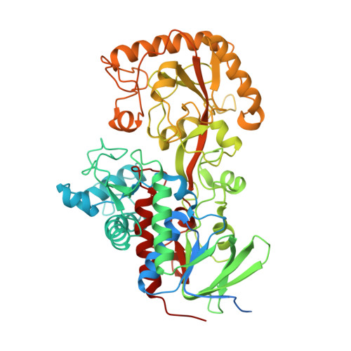
    The selective α,β-desaturation of cyclic carbonyl compounds, which are found in the core of many steroid and bioactive molecules, using green chemistry is highly desirable. To achieve this task, we have for the first time described and solved the de novo structure of a member of the cyclohexanone dehydrogenase class of enzymes. The breadth of substrate specificity was investigated by assaying the cyclohexanone dehydrogenase, from Alicycliphilus denitrificans , against several cyclic ketones, lactones and lactams. To investigate substrate binding, a catalytic variant, Y195F, was generated and used to obtain a crystallographic complex with the natural substrate, cyclohexanone. This revealed substrate-active site interactions, as well as the proximity of the cofactor, flavin adenine dinucleotide, and enabled us to propose a mechanistic function to key amino acids. We then used molecular dynamic simulations to guide design to add functionality to the cyclohexanone dehydrogenase enzyme. The resulting W113A variant had overall improved enzyme activity and substrate scope, i.e. , accepting the bulkier carbonyl compound, dihydrocoumarin. Structural analysis of the W113A variant revealed a broader, more open active site, which helped explain the modified substrate specificity. This work paves the way for future bespoke regioselective α,β-desaturation in the synthesis of important bioactive molecules via rational enzyme engineering.


  • Organizational Affiliation
    • Hub for Biotechnology in Build Environment, Department of Applied Sciences, Faculty of Health and Life Sciences, Northumbria University Newcastle upon Tyne NE1 8ST UK gary.black@northumbria.ac.uk.

Macromolecule Content 

  • Total Structure Weight: 131.09 kDa 
  • Atom Count: 9,446 
  • Modeled Residue Count: 1,072 
  • Deposited Residue Count: 1,196 
  • Unique protein chains: 1

Macromolecules

Find similar proteins by:|  3D Structure
Entity ID: 1
MoleculeChains  Sequence LengthOrganismDetailsImage
Fumarate reductase/succinate dehydrogenase flavoprotein domain proteinA [auth AAA],
B [auth BBB]
598Alicycliphilus denitrificans K601Mutation(s): 0 
Gene Names: Alide2_4318
UniProt
Find proteins for F4G7N3 (Alicycliphilus denitrificans (strain DSM 14773 / CIP 107495 / K601))
Explore F4G7N3 
Go to UniProtKB:  F4G7N3
Entity Groups
Sequence Clusters30% Identity50% Identity70% Identity90% Identity95% Identity100% Identity
UniProt GroupF4G7N3
Sequence Annotations
Expand
Reference Sequence

Small Molecules

Ligands 3 Unique
IDChains Name / Formula / InChI Key2D Diagram3D Interactions
FAD
(Subject of Investigation/LOI)

Query on FAD


Download:Ideal Coordinates CCD File
C [auth AAA],
G [auth BBB]
FLAVIN-ADENINE DINUCLEOTIDE
C27 H33 N9 O15 P2
VWWQXMAJTJZDQX-UYBVJOGSSA-N
SO4

Query on SO4


Download:Ideal Coordinates CCD File
E [auth AAA],
F [auth AAA],
H [auth BBB]
SULFATE ION
O4 S
QAOWNCQODCNURD-UHFFFAOYSA-L
GOL

Query on GOL


Download:Ideal Coordinates CCD File
D [auth AAA]GLYCEROL
C3 H8 O3
PEDCQBHIVMGVHV-UHFFFAOYSA-N

Experimental Data & Validation

Experimental Data

  • Method: X-RAY DIFFRACTION
  • Resolution: 1.86 Å
  • R-Value Free:  0.207 (Depositor) 
  • R-Value Work:  0.171 (Depositor) 
Space Group: P 43 21 2
Unit Cell:
Length ( Å )Angle ( ˚ )
a = 90.699α = 90
b = 90.699β = 90
c = 278.239γ = 90
Software Package:
Software NamePurpose
REFMACrefinement
PHASERphasing
SHELXDEphasing
Cootmodel building
CRANKphasing
PDB_EXTRACTdata extraction

Structure Validation

View Full Validation Report


Ligand Structure Quality Assessment 


Entry History 

& Funding Information

Deposition Data


Funding OrganizationLocationGrant Number
Biotechnology and Biological Sciences Research Council (BBSRC)United Kingdom1065600

Revision History  (Full details and data files)

  • Version 1.0: 2024-02-14
    Type: Initial release
  • Version 1.1: 2024-04-10
    Changes: Database references