8AM0

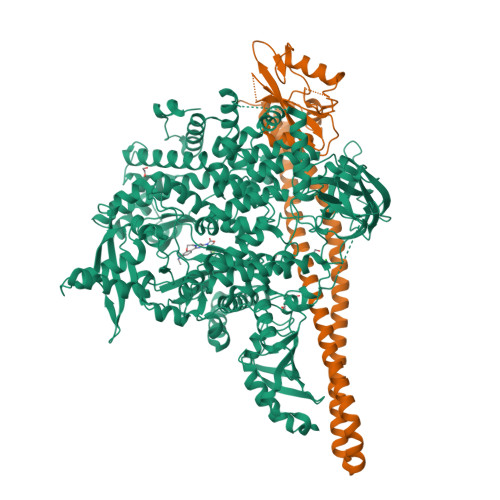
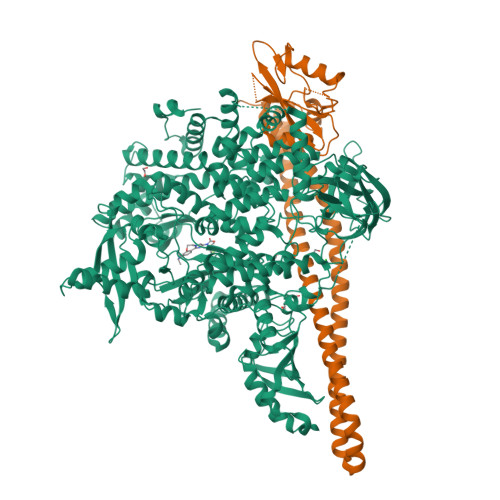
Crystal structure of human T1061E PI3Kalpha in complex with its regulatory subunit and the inhibitor GDC-0077 (Inavolisib)


Experimental Data Snapshot

  • Method: X-RAY DIFFRACTION
  • Resolution: 2.82 Å
  • R-Value Free: 0.300 
  • R-Value Work: 0.285 
  • R-Value Observed: 0.286 

wwPDB Validation   3D Report Full Report


Ligand Structure Quality Assessment 


This is version 1.1 of the entry. See complete history


Literature

Epinephrine inhibits PI3K alpha via the Hippo kinases.

Lin, T.Y.Ramsamooj, S.Perrier, T.Liberatore, K.Lantier, L.Vasan, N.Karukurichi, K.Hwang, S.K.Kesicki, E.A.Kastenhuber, E.R.Wiederhold, T.Yaron, T.M.Huntsman, E.M.Zhu, M.Ma, Y.Paddock, M.N.Zhang, G.Hopkins, B.D.McGuinness, O.Schwartz, R.E.Ersoy, B.A.Cantley, L.C.Johnson, J.L.Goncalves, M.D.

(2023) Cell Rep 42: 113535-113535

  • DOI: https://doi.org/10.1016/j.celrep.2023.113535
  • Primary Citation of Related Structures:  
    8AM0

  • PubMed Abstract: 

    The phosphoinositide 3-kinase p110α is an essential mediator of insulin signaling and glucose homeostasis. We interrogated the human serine, threonine, and tyrosine kinome to search for novel regulators of p110α and found that the Hippo kinases phosphorylate p110α at T1061, which inhibits its activity. This inhibitory state corresponds to a conformational change of a membrane-binding domain on p110α, which impairs its ability to engage membranes. In human primary hepatocytes, cancer cell lines, and rodent tissues, activation of the Hippo kinases MST1/2 using forskolin or epinephrine is associated with phosphorylation of T1061 and inhibition of p110α, impairment of downstream insulin signaling, and suppression of glycolysis and glycogen synthesis. These changes are abrogated when MST1/2 are genetically deleted or inhibited with small molecules or if the T1061 is mutated to alanine. Our study defines an inhibitory pathway of PI3K signaling and a link between epinephrine and insulin signaling.


  • Organizational Affiliation

    Meyer Cancer Center, Weill Cornell Medicine, New York, NY 10021, USA; Weill Cornell Graduate School of Medical Sciences, New York, NY 10021, USA.


Macromolecules
Find similar proteins by:  (by identity cutoff)  |  3D Structure
Entity ID: 1
MoleculeChains Sequence LengthOrganismDetailsImage
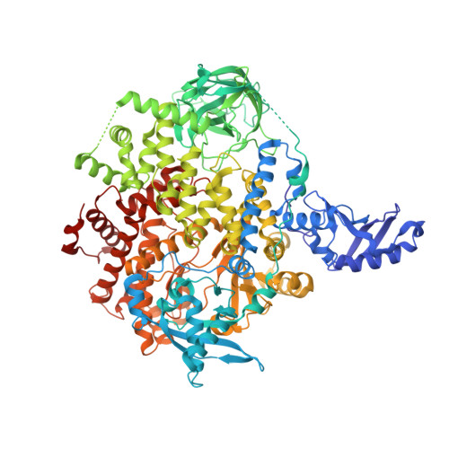
Phosphatidylinositol 4,5-bisphosphate 3-kinase catalytic subunit alpha isoform1,068Homo sapiensMutation(s): 1 
Gene Names: PIK3CA
EC: 2.7.1.137 (PDB Primary Data), 2.7.1.153 (PDB Primary Data), 2.7.11.1 (PDB Primary Data)
UniProt & NIH Common Fund Data Resources
Find proteins for P42336 (Homo sapiens)
Explore P42336 
Go to UniProtKB:  P42336
PHAROS:  P42336
GTEx:  ENSG00000121879 
Entity Groups  
Sequence Clusters30% Identity50% Identity70% Identity90% Identity95% Identity100% Identity
UniProt GroupP42336
Sequence Annotations
Expand
  • Reference Sequence
Find similar proteins by:  (by identity cutoff)  |  3D Structure
Entity ID: 2
MoleculeChains Sequence LengthOrganismDetailsImage
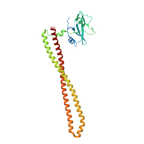
Isoform 3 of Phosphatidylinositol 3-kinase regulatory subunit alpha293Homo sapiensMutation(s): 0 
Gene Names: PIK3R1GRB1
UniProt & NIH Common Fund Data Resources
Find proteins for P27986 (Homo sapiens)
Explore P27986 
Go to UniProtKB:  P27986
PHAROS:  P27986
GTEx:  ENSG00000145675 
Entity Groups  
Sequence Clusters30% Identity50% Identity70% Identity90% Identity95% Identity100% Identity
UniProt GroupP27986
Sequence Annotations
Expand
  • Reference Sequence
Small Molecules
Ligands 4 Unique
IDChains Name / Formula / InChI Key2D Diagram3D Interactions
MWF (Subject of Investigation/LOI)
Query on MWF

Download Ideal Coordinates CCD File 
C [auth A](2R)-2-[[2-[(4S)-4-[bis(fluoranyl)methyl]-2-oxidanylidene-1,3-oxazolidin-3-yl]-5,6-dihydroimidazo[1,2-d][1,4]benzoxazepin-9-yl]amino]propanamide
C18 H19 F2 N5 O4
SGEUNORSOZVTOL-SKDRFNHKSA-N
PGE
Query on PGE

Download Ideal Coordinates CCD File 
F [auth A]TRIETHYLENE GLYCOL
C6 H14 O4
ZIBGPFATKBEMQZ-UHFFFAOYSA-N
EDO
Query on EDO

Download Ideal Coordinates CCD File 
D [auth A],
E [auth A],
H [auth B]
1,2-ETHANEDIOL
C2 H6 O2
LYCAIKOWRPUZTN-UHFFFAOYSA-N
CL
Query on CL

Download Ideal Coordinates CCD File 
G [auth A]CHLORIDE ION
Cl
VEXZGXHMUGYJMC-UHFFFAOYSA-M
Experimental Data & Validation

Experimental Data

  • Method: X-RAY DIFFRACTION
  • Resolution: 2.82 Å
  • R-Value Free: 0.300 
  • R-Value Work: 0.285 
  • R-Value Observed: 0.286 
  • Space Group: P 21 21 21
Unit Cell:
Length ( Å )Angle ( ˚ )
a = 99.81α = 90
b = 107.68β = 90
c = 136.3γ = 90
Software Package:
Software NamePurpose
BUSTERrefinement
XDSdata reduction
STARANISOdata scaling
PHASERphasing

Structure Validation

View Full Validation Report


Ligand Structure Quality Assessment 


Entry History & Funding Information

Deposition Data


Funding OrganizationLocationGrant Number
National Institutes of Health/National Cancer Institute (NIH/NCI)United StatesR35 CA197588

Revision History  (Full details and data files)

  • Version 1.0: 2023-12-13
    Type: Initial release
  • Version 1.1: 2024-01-03
    Changes: Database references