8AEU | pdb_00008aeu

Structure of hMDM2 in complex with Nutlin-3a-aa


Experimental Data Snapshot

  • Method: X-RAY DIFFRACTION
  • Resolution: 2.00 Å
  • R-Value Free: 
    0.240 (Depositor), 0.234 (DCC) 
  • R-Value Work: 
    0.197 (Depositor), 0.219 (DCC) 
  • R-Value Observed: 
    0.201 (Depositor) 

Starting Model: experimental
View more details

wwPDB Validation 3D Report Full Report

Validation slider image for 8AEU

Ligand Structure Quality Assessment 


This is version 1.4 of the entry. See complete history

Literature

Nutlin-3a-aa: Improving the Bioactivity of a p53/MDM2 Interaction Inhibitor by Introducing a Solvent-Exposed Methylene Group.

Nietzold, F.Rubner, S.Labuzek, B.Golik, P.Surmiak, E.Del Corte, X.Kitel, R.Protzel, C.Reppich-Sacher, R.Stichel, J.Magiera-Mularz, K.Holak, T.A.Berg, T.

(2023) Chembiochem 24: e202300006-e202300006

  • DOI: https://doi.org/10.1002/cbic.202300006
  • Primary Citation Related Structures: 
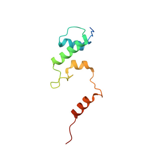
    8AEU

  • PubMed Abstract: 

    Nutlin-3a is a reversible inhibitor of the p53/MDM2 interaction. We have synthesized the derivative Nutlin-3a-aa bearing an additional exocyclic methylene group in the piperazinone moiety. Nutlin-3a-aa is more active than Nutlin-3a against purified wild-type MDM2, and is more effective at increasing p53 levels and releasing transcription of p53 target genes from MDM2-induced repression. X-ray analysis of wild-type MDM2-bound Nutlin-3a-aa indicated that the orientation of its modified piperazinone ring was altered in comparison to the piperazinone ring of MDM2-bound Nutlin-3a, with the exocyclic methylene group of Nutlin-3a-aa pointing away from the protein surface. Our data point to the introduction of exocyclic methylene groups as a useful approach by which to tailor the conformation of bioactive molecules for improved biological activity.


  • Organizational Affiliation
    • Institute of Organic Chemistry, Leipzig University, Johannisallee 29, 04103, Leipzig, Germany.

Macromolecule Content 

  • Total Structure Weight: 12.07 kDa 
  • Atom Count: 777 
  • Modeled Residue Count: 87 
  • Deposited Residue Count: 97 
  • Unique protein chains: 1

Macromolecules

Find similar proteins by:|  3D Structure
Entity ID: 1
MoleculeChains  Sequence LengthOrganismDetailsImage
E3 ubiquitin-protein ligase Mdm297Homo sapiensMutation(s): 0 
EC: 2.3.2.27
UniProt & NIH Common Fund Data Resources
Find proteins for Q00987 (Homo sapiens)
Explore Q00987 
Go to UniProtKB:  Q00987
PHAROS:  Q00987
GTEx:  ENSG00000135679 
Entity Groups
Sequence Clusters30% Identity50% Identity70% Identity90% Identity95% Identity100% Identity
UniProt GroupQ00987
Sequence Annotations
Expand
Reference Sequence

Small Molecules

Ligands 2 Unique
IDChains Name / Formula / InChI Key2D Diagram3D Interactions
M0L
(Subject of Investigation/LOI)

Query on M0L


Download:Ideal Coordinates CCD File
C [auth A]4-[[(4~{S},5~{R})-4,5-bis(4-chlorophenyl)-2-(4-methoxy-2-propan-2-yloxy-phenyl)-4,5-dihydroimidazol-1-yl]carbonyl]-3-methylidene-piperazin-2-one
C31 H30 Cl2 N4 O4
JLTUTZCZAIJXTD-WUFINQPMSA-N
BME

Query on BME


Download:Ideal Coordinates CCD File
B [auth A]BETA-MERCAPTOETHANOL
C2 H6 O S
DGVVWUTYPXICAM-UHFFFAOYSA-N

Experimental Data & Validation

Experimental Data

  • Method: X-RAY DIFFRACTION
  • Resolution: 2.00 Å
  • R-Value Free:  0.240 (Depositor), 0.234 (DCC) 
  • R-Value Work:  0.197 (Depositor), 0.219 (DCC) 
  • R-Value Observed: 0.201 (Depositor) 
Space Group: P 43 21 2
Unit Cell:
Length ( Å )Angle ( ˚ )
a = 56.233α = 90
b = 56.233β = 90
c = 75.376γ = 90
Software Package:
Software NamePurpose
PHENIXrefinement
PDB_EXTRACTdata extraction
XDSdata reduction
Aimlessdata scaling
PHASERphasing

Structure Validation

View Full Validation Report


Ligand Structure Quality Assessment 


Entry History 

& Funding Information

Deposition Data


Funding OrganizationLocationGrant Number
Polish National Science CentrePoland2014/12/W/NZ1/00457

Revision History  (Full details and data files)

  • Version 1.0: 2023-01-18
    Type: Initial release
  • Version 1.1: 2023-03-01
    Changes: Database references
  • Version 1.2: 2023-03-22
    Changes: Database references
  • Version 1.3: 2023-03-29
    Changes: Database references
  • Version 1.4: 2024-02-07
    Changes: Data collection, Refinement description