8A9N | pdb_00008a9n

Structure of DpA polyamine acetyltransferase in complex with 1,3-DAP


Experimental Data Snapshot

  • Method: X-RAY DIFFRACTION
  • Resolution: 1.85 Å
  • R-Value Free: 
    0.251 (Depositor), 0.244 (DCC) 
  • R-Value Work: 
    0.206 (Depositor), 0.203 (DCC) 
  • R-Value Observed: 
    0.208 (Depositor) 

Starting Model: experimental
View more details

wwPDB Validation 3D Report Full Report

Validation slider image for 8A9N

Ligand Structure Quality Assessment 


This is version 1.2 of the entry. See complete history

Literature

A polyamine acetyltransferase regulates the motility and biofilm formation of Acinetobacter baumannii.

Armalyte, J.Cepauskas, A.Sakalyte, G.Martinkus, J.Skerniskyte, J.Martens, C.Suziedeliene, E.Garcia-Pino, A.Jurenas, D.

(2023) Nat Commun 14: 3531-3531

  • DOI: https://doi.org/10.1038/s41467-023-39316-5
  • Primary Citation Related Structures: 
    8A9N, 8A9O

  • PubMed Abstract: 

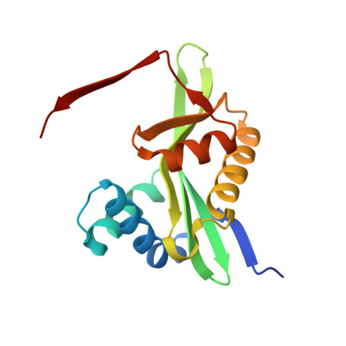
    Acinetobacter baumannii is a nosocomial pathogen highly resistant to environmental changes and antimicrobial treatments. Regulation of cellular motility and biofilm formation is important for its virulence, although it is poorly described at the molecular level. It has been previously reported that Acinetobacter genus specifically produces a small positively charged metabolite, polyamine 1,3-diaminopropane, that has been associated with cell motility and virulence. Here we show that A. baumannii encodes novel acetyltransferase, Dpa, that acetylates 1,3-diaminopropane, directly affecting the bacterium motility. Expression of dpa increases in bacteria that form pellicle and adhere to eukaryotic cells as compared to planktonic bacterial cells, suggesting that cell motility is linked to the pool of non-modified 1,3-diaminopropane. Indeed, deletion of dpa hinders biofilm formation and increases twitching motion confirming the impact of balancing the levels of 1,3-diaminopropane on cell motility. The crystal structure of Dpa reveals topological and functional differences from other bacterial polyamine acetyltransferases, adopting a β-swapped quaternary arrangement similar to that of eukaryotic polyamine acetyltransferases with a central size exclusion channel that sieves through the cellular polyamine pool. The structure of catalytically impaired Dpa Y128F in complex with the reaction product shows that binding and orientation of the polyamine substrates are conserved between different polyamine-acetyltransferases.


  • Organizational Affiliation
    • Institute of Biosciences, Life Sciences Center, Vilnius University, Saulėtekio av. 7, LT-10257, Vilnius, Lithuania.

Macromolecule Content 

  • Total Structure Weight: 38.54 kDa 
  • Atom Count: 2,883 
  • Modeled Residue Count: 299 
  • Deposited Residue Count: 308 
  • Unique protein chains: 1

Macromolecules

Find similar proteins by:|  3D Structure
Entity ID: 1
MoleculeChains  Sequence LengthOrganismDetailsImage
Acetyltransferase
A, B
154Acinetobacter baumanniiMutation(s): 0 
Gene Names: 
UniProt
Find proteins for V5VBK4 (Acinetobacter baumannii)
Explore V5VBK4 
Go to UniProtKB:  V5VBK4
Entity Groups
Sequence Clusters30% Identity50% Identity70% Identity90% Identity95% Identity100% Identity
UniProt GroupV5VBK4
Sequence Annotations
Expand
Reference Sequence

Small Molecules

Ligands 5 Unique
IDChains Name / Formula / InChI Key2D Diagram3D Interactions
COA
(Subject of Investigation/LOI)

Query on COA


Download:Ideal Coordinates CCD File
C [auth A],
G [auth B]
COENZYME A
C21 H36 N7 O16 P3 S
RGJOEKWQDUBAIZ-IBOSZNHHSA-N
LFU

Query on LFU


Download:Ideal Coordinates CCD File
E [auth A]~{N}-(3-azanylpropyl)ethanamide
C5 H12 N2 O
YFZBPSXRYCOKCW-UHFFFAOYSA-N
GOL

Query on GOL


Download:Ideal Coordinates CCD File
F [auth B]GLYCEROL
C3 H8 O3
PEDCQBHIVMGVHV-UHFFFAOYSA-N
13D

Query on 13D


Download:Ideal Coordinates CCD File
I [auth B]1,3-DIAMINOPROPANE
C3 H10 N2
XFNJVJPLKCPIBV-UHFFFAOYSA-N
MG

Query on MG


Download:Ideal Coordinates CCD File
D [auth A],
H [auth B]
MAGNESIUM ION
Mg
JLVVSXFLKOJNIY-UHFFFAOYSA-N

Experimental Data & Validation

Experimental Data

  • Method: X-RAY DIFFRACTION
  • Resolution: 1.85 Å
  • R-Value Free:  0.251 (Depositor), 0.244 (DCC) 
  • R-Value Work:  0.206 (Depositor), 0.203 (DCC) 
  • R-Value Observed: 0.208 (Depositor) 
Space Group: P 1 21 1
Unit Cell:
Length ( Å )Angle ( ˚ )
a = 42.87α = 90
b = 68.08β = 109.12
c = 55.74γ = 90
Software Package:
Software NamePurpose
BUSTERrefinement
XDSdata reduction
Aimlessdata scaling
PHASERphasing

Structure Validation

View Full Validation Report


Ligand Structure Quality Assessment 


Entry History 

& Funding Information

Deposition Data


Funding OrganizationLocationGrant Number
Fonds National de la Recherche Scientifique (FNRS)BelgiumT.0090.22

Revision History  (Full details and data files)

  • Version 1.0: 2023-07-12
    Type: Initial release
  • Version 1.1: 2023-11-22
    Changes: Data collection, Database references
  • Version 1.2: 2024-02-07
    Changes: Refinement description