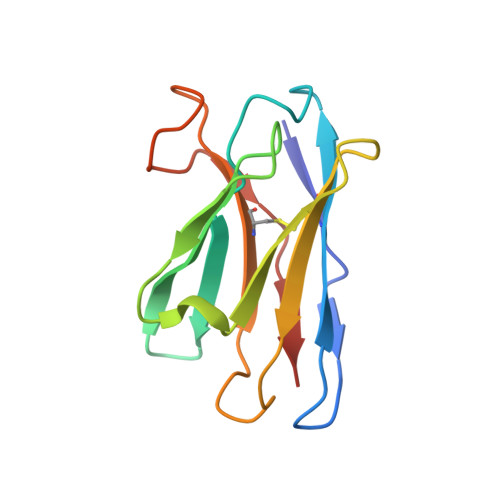
8A1E | pdb_00008a1e

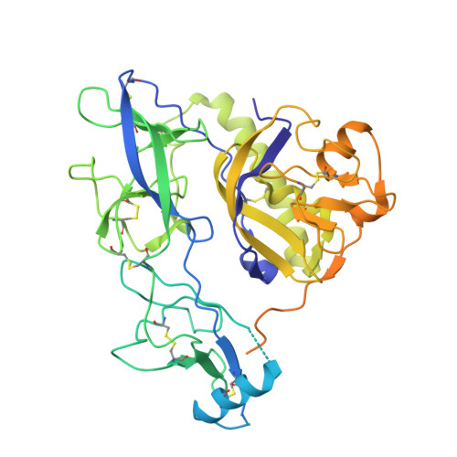
Rabies virus glycoprotein in complex with Fab fragments of 17C7 and 1112-1 neutralizing antibodies


Experimental Data Snapshot

  • Method: ELECTRON MICROSCOPY
  • Resolution: 2.83 Å
  • Aggregation State: PARTICLE 
  • Reconstruction Method: SINGLE PARTICLE 

wwPDB Validation 3D Report Full Report

Validation slider image for 8A1E

This is version 1.3 of the entry. See complete history. 

Literature

Structure of trimeric pre-fusion rabies virus glycoprotein in complex with two protective antibodies.

Ng, W.M., Fedosyuk, S., English, S., Augusto, G., Berg, A., Thorley, L., Haselon, A.S., Segireddy, R.R., Bowden, T.A., Douglas, A.D.

(2022) Cell Host Microbe 30: 1219-1230.e7

  • DOI: https://doi.org/10.1016/j.chom.2022.07.014
  • Primary Citation Related Structures: 
    8A1E

  • PubMed Abstract: 

    Rabies virus (RABV) causes lethal encephalitis and is responsible for approximately 60,000 deaths per year. As the sole virion-surface protein, the rabies virus glycoprotein (RABV-G) mediates host-cell entry. RABV-G's pre-fusion trimeric conformation displays epitopes bound by protective neutralizing antibodies that can be induced by vaccination or passively administered for post-exposure prophylaxis. We report a 2.8-Å structure of a RABV-G trimer in the pre-fusion conformation, in complex with two neutralizing and protective monoclonal antibodies, 17C7 and 1112-1, that recognize distinct epitopes. One of these antibodies is a licensed prophylactic (17C7, Rabishield), which we show locks the protein in pre-fusion conformation. Targeted mutations can similarly stabilize RABV-G in the pre-fusion conformation, a key step toward structure-guided vaccine design. These data reveal the higher-order architecture of a key therapeutic target and the structural basis of neutralization by antibodies binding two key antigenic sites, and this will facilitate the development of improved vaccines and prophylactic antibodies.


  • Organizational Affiliation: 
    • Jenner Institute, Old Road Campus Research Building, Roosevelt Drive, Oxford OX3 7DQ, UK; Division of Structural Biology, Wellcome Centre for Human Genetics, University of Oxford, Roosevelt Drive, Oxford OX3 7BN, UK.

Macromolecule Content 

  • Total Structure Weight: 106.57 kDa 
  • Atom Count: 6,134 
  • Modeled Residue Count: 790 
  • Deposited Residue Count: 961 
  • Unique protein chains: 5

Macromolecules

Find similar proteins by:|  3D Structure
Entity ID: 1
MoleculeChains  Sequence LengthOrganismDetailsImage
Glycoprotein505Rabies virus strain Pasteur vaccinMutation(s): 0 
UniProt
Find proteins for P08667 (Rabies virus (strain Pasteur vaccins / PV))
Explore P08667 
Go to UniProtKB:  P08667
Entity Groups
Sequence Clusters30% Identity50% Identity70% Identity90% Identity95% Identity100% Identity
UniProt GroupP08667
Sequence Annotations
Expand
Reference Sequence
Find similar proteins by:|  3D Structure
Entity ID: 2
MoleculeChains  Sequence LengthOrganismDetailsImage
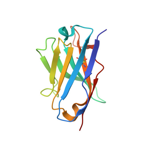
Fab 17C7 heavy chain variable domainB [auth D]119Homo sapiensMutation(s): 0 
Entity Groups
Sequence Clusters30% Identity50% Identity70% Identity90% Identity95% Identity100% Identity
Sequence Annotations
Expand
Reference Sequence
Find similar proteins by:|  3D Structure
Entity ID: 3
MoleculeChains  Sequence LengthOrganismDetailsImage
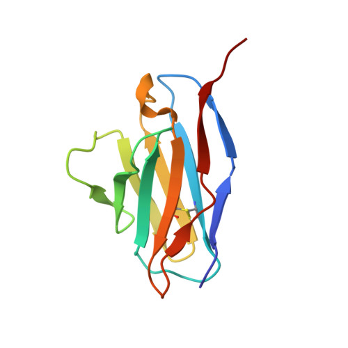
Fab 17C7 light chain variable domainC [auth E]109Homo sapiensMutation(s): 0 
Entity Groups
Sequence Clusters30% Identity50% Identity70% Identity90% Identity95% Identity100% Identity
Sequence Annotations
Expand
Reference Sequence
Find similar proteins by:|  3D Structure
Entity ID: 4
MoleculeChains  Sequence LengthOrganismDetailsImage
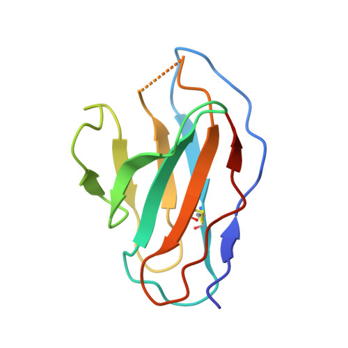
Fab 1112-1 light chain variable domainD [auth C]107Mus musculusMutation(s): 0 
Entity Groups
Sequence Clusters30% Identity50% Identity70% Identity90% Identity95% Identity100% Identity
Sequence Annotations
Expand
Reference Sequence
Find similar proteins by:|  3D Structure
Entity ID: 5
MoleculeChains  Sequence LengthOrganismDetailsImage
Fab 1112-1 heavy chain variable domainE [auth B]121Mus musculusMutation(s): 0 
Entity Groups
Sequence Clusters30% Identity50% Identity70% Identity90% Identity95% Identity100% Identity
Sequence Annotations
Expand
Reference Sequence

Experimental Data & Validation

Experimental Data

  • Method: ELECTRON MICROSCOPY
  • Resolution: 2.83 Å
  • Aggregation State: PARTICLE 
  • Reconstruction Method: SINGLE PARTICLE 
EM Software:
TaskSoftware PackageVersion
RECONSTRUCTIONcryoSPARC3.1
MODEL REFINEMENTPHENIX1.19.2_4158

Structure Validation

View Full Validation Report


Entry History 

& Funding Information

Deposition Data


Funding OrganizationLocationGrant Number
Wellcome TrustUnited Kingdom220679/Z/20/Z
Medical Research Council (MRC, United Kingdom)United KingdomMR/P017339/1
Medical Research Council (MRC, United Kingdom)United KingdomMR/S007555/1
Marie Sklodowska-Curie Actions, FragNET ITNEuropean Union840866
Wellcome TrustUnited Kingdom060208/Z/00/Z
Wellcome TrustUnited Kingdom093305/Z/10/Z
Wellcome TrustUnited Kingdom203141/Z/16/Z

Revision History  (Full details and data files)

  • Version 1.0: 2022-08-17
    Type: Initial release
  • Version 1.1: 2022-08-31
    Changes: Database references
  • Version 1.2: 2022-09-28
    Changes: Database references
  • Version 1.3: 2024-11-06
    Changes: Data collection, Structure summary