7ZZL

Crystal structure of CYP106A1


Experimental Data Snapshot

  • Method: X-RAY DIFFRACTION
  • Resolution: 1.70 Å
  • R-Value Free: 0.206 
  • R-Value Work: 0.169 
  • R-Value Observed: 0.171 

wwPDB Validation   3D Report Full Report


Ligand Structure Quality Assessment 


This is version 1.1 of the entry. See complete history


Literature

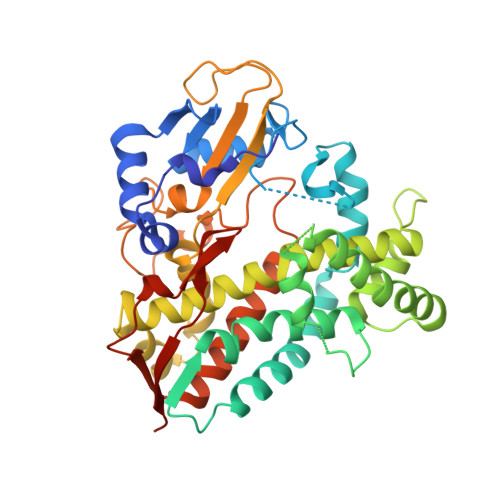
Structural comparison of the cytochrome P450 enzymes CYP106A1 and CYP106A2 provides insight into their differences in steroid conversion.

Carius, Y.Hutter, M.Kiss, F.Bernhardt, R.Lancaster, C.R.D.

(2022) FEBS Lett 596: 3133-3144

  • DOI: https://doi.org/10.1002/1873-3468.14502
  • Primary Citation of Related Structures:  
    7Q9E, 7ZZL

  • PubMed Abstract: 

    Understanding the structural basis of the selectivity of steroid hydroxylation requires detailed structural and functional investigations on various steroid hydroxylases with different selectivities, such as the bacterial cytochrome P450 enzymes. Here, the crystal structure of the cytochrome P450 CYP106A1 from Priestia megaterium was solved. CYP106A1 exhibits a rare additional structural motif of a cytochrome P450, a sixth β-sheet. The protein was found in different unusual conformations corresponding to both open and closed forms even when crystallized without any known substrate. The structural comparison of CYP106A1 with the previously investigated CYP106A2, including docking studies for both isoforms with the substrate cortisol, reveals a completely different orientation of the steroid molecule in the active sites. This distinction convincingly explains the experimentally observed differences in substrate conversion and product formation by the two enzymes.


  • Organizational Affiliation

    Department of Structural Biology, Faculty of Medicine, Center of Human and Molecular Biology (ZHMB), Saarland University, Homburg, Germany.


Macromolecules
Find similar proteins by:  (by identity cutoff)  |  3D Structure
Entity ID: 1
MoleculeChains Sequence LengthOrganismDetailsImage
Cytochrome P450
A, B, C, D
416Priestia megaterium DSM 319Mutation(s): 0 
Gene Names: BMD_1855
EC: 1.14.14
UniProt
Find proteins for D5DF35 (Priestia megaterium (strain DSM 319 / IMG 1521))
Explore D5DF35 
Go to UniProtKB:  D5DF35
Entity Groups  
Sequence Clusters30% Identity50% Identity70% Identity90% Identity95% Identity100% Identity
UniProt GroupD5DF35
Sequence Annotations
Expand
  • Reference Sequence
Small Molecules
Ligands 7 Unique
IDChains Name / Formula / InChI Key2D Diagram3D Interactions
HEM (Subject of Investigation/LOI)
Query on HEM

Download Ideal Coordinates CCD File 
DA [auth D],
E [auth A],
L [auth B],
V [auth C]
PROTOPORPHYRIN IX CONTAINING FE
C34 H32 Fe N4 O4
KABFMIBPWCXCRK-RGGAHWMASA-L
PEG
Query on PEG

Download Ideal Coordinates CCD File 
Q [auth B],
Z [auth C]
DI(HYDROXYETHYL)ETHER
C4 H10 O3
MTHSVFCYNBDYFN-UHFFFAOYSA-N
SO4
Query on SO4

Download Ideal Coordinates CCD File 
AA [auth C]
BA [auth C]
EA [auth D]
F [auth A]
FA [auth D]
AA [auth C],
BA [auth C],
EA [auth D],
F [auth A],
FA [auth D],
G [auth A],
H [auth A],
I [auth A],
J [auth A],
K [auth A],
M [auth B],
N [auth B],
P [auth B],
T [auth B],
U [auth B],
W [auth C],
X [auth C]
SULFATE ION
O4 S
QAOWNCQODCNURD-UHFFFAOYSA-L
DMS
Query on DMS

Download Ideal Coordinates CCD File 
O [auth B]DIMETHYL SULFOXIDE
C2 H6 O S
IAZDPXIOMUYVGZ-UHFFFAOYSA-N
IMD
Query on IMD

Download Ideal Coordinates CCD File 
Y [auth C]IMIDAZOLE
C3 H5 N2
RAXXELZNTBOGNW-UHFFFAOYSA-O
CO
Query on CO

Download Ideal Coordinates CCD File 
GA [auth D],
R [auth B],
S [auth B]
COBALT (II) ION
Co
XLJKHNWPARRRJB-UHFFFAOYSA-N
OXY
Query on OXY

Download Ideal Coordinates CCD File 
CA [auth C]OXYGEN MOLECULE
O2
MYMOFIZGZYHOMD-UHFFFAOYSA-N
Experimental Data & Validation

Experimental Data

  • Method: X-RAY DIFFRACTION
  • Resolution: 1.70 Å
  • R-Value Free: 0.206 
  • R-Value Work: 0.169 
  • R-Value Observed: 0.171 
  • Space Group: P 1
Unit Cell:
Length ( Å )Angle ( ˚ )
a = 63.69α = 95.36
b = 82.87β = 90
c = 84.86γ = 90
Software Package:
Software NamePurpose
REFMACrefinement
PDB_EXTRACTdata extraction
MOSFLMdata reduction
SCALAdata scaling
MOLREPphasing

Structure Validation

View Full Validation Report


Ligand Structure Quality Assessment 


Entry History & Funding Information

Deposition Data


Funding OrganizationLocationGrant Number
Other government--

Revision History  (Full details and data files)

  • Version 1.0: 2023-04-12
    Type: Initial release
  • Version 1.1: 2024-02-07
    Changes: Data collection, Refinement description