7ZH1 | pdb_00007zh1

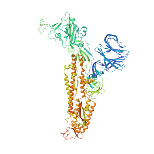
SARS CoV Spike protein, Closed C3 conformation


Experimental Data Snapshot

  • Method: ELECTRON MICROSCOPY
  • Resolution: 2.48 Å
  • Aggregation State: PARTICLE 
  • Reconstruction Method: SINGLE PARTICLE 

Starting Model: experimental
View more details

wwPDB Validation 3D Report Full Report

Validation slider image for 7ZH1

This is version 1.1 of the entry. See complete history. 

Literature

The free fatty acid-binding pocket is a conserved hallmark in pathogenic beta-coronavirus spike proteins from SARS-CoV to Omicron.

Toelzer, C., Gupta, K., Yadav, S.K.N., Hodgson, L., Williamson, M.K., Buzas, D., Borucu, U., Powers, K., Stenner, R., Vasileiou, K., Garzoni, F., Fitzgerald, D., Payre, C., Gautam, G., Lambeau, G., Davidson, A.D., Verkade, P., Frank, M., Berger, I., Schaffitzel, C.

(2022) Sci Adv 8: eadc9179-eadc9179

  • DOI: https://doi.org/10.1126/sciadv.adc9179
  • Primary Citation Related Structures: 
    7ZH1, 7ZH2, 7ZH5

  • PubMed Abstract: 

    As coronavirus disease 2019 (COVID-19) persists, severe acute respiratory syndrome coronavirus 2 (SARS-CoV-2) variants of concern (VOCs) emerge, accumulating spike (S) glycoprotein mutations. S receptor binding domain (RBD) comprises a free fatty acid (FFA)-binding pocket. FFA binding stabilizes a locked S conformation, interfering with virus infectivity. We provide evidence that the pocket is conserved in pathogenic β-coronaviruses (β-CoVs) infecting humans. SARS-CoV, MERS-CoV, SARS-CoV-2, and VOCs bind the essential FFA linoleic acid (LA), while binding is abolished by one mutation in common cold-causing HCoV-HKU1. In the SARS-CoV S structure, LA stabilizes the locked conformation, while the open, infectious conformation is devoid of LA. Electron tomography of SARS-CoV-2-infected cells reveals that LA treatment inhibits viral replication, resulting in fewer deformed virions. Our results establish FFA binding as a hallmark of pathogenic β-CoV infection and replication, setting the stage for FFA-based antiviral strategies to overcome COVID-19.


  • Organizational Affiliation: 
    • School of Biochemistry, University of Bristol, 1 Tankard's Close, Bristol BS8 1TD, UK.

Macromolecule Content 

  • Total Structure Weight: 423.36 kDa 
  • Atom Count: 24,501 
  • Modeled Residue Count: 3,060 
  • Deposited Residue Count: 3,741 
  • Unique protein chains: 1

Macromolecules

Find similar proteins by:|  3D Structure
Entity ID: 1
MoleculeChains  Sequence LengthOrganismDetailsImage
Spike glycoprotein,Fibritin
A, B, C
1,247Severe acute respiratory syndrome-related coronavirus, Tequatrovirus T4
This entity is chimeric
Mutation(s): 2 
Gene Names: S, 2, wac
UniProt
Find proteins for P10104 (Enterobacteria phage T4)
Explore P10104 
Go to UniProtKB:  P10104
Find proteins for P59594 (Severe acute respiratory syndrome coronavirus)
Explore P59594 
Go to UniProtKB:  P59594
Entity Groups
Sequence Clusters30% Identity50% Identity70% Identity90% Identity95% Identity100% Identity
UniProt GroupsP59594P10104
Glycosylation
Glycosylation Sites: 9Go to GlyGen: P59594-1
Sequence Annotations
Expand
Reference Sequence

Oligosaccharides

Help  
Entity ID: 2
MoleculeChains Length2D Diagram GlycosylationD Interactions
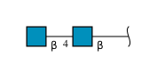
2-acetamido-2-deoxy-beta-D-glucopyranose-(1-4)-2-acetamido-2-deoxy-beta-D-glucopyranose
D, E, F
2N-Glycosylation
Glycosylation Resources
GlyTouCan: G42666HT
GlyCosmos: G42666HT
GlyGen: G42666HT

Small Molecules

Ligands 2 Unique
IDChains Name / Formula / InChI Key2D Diagram3D Interactions
EIC
(Subject of Investigation/LOI)

Query on EIC


Download:Ideal Coordinates CCD File
CA [auth C],
G [auth A],
R [auth B]
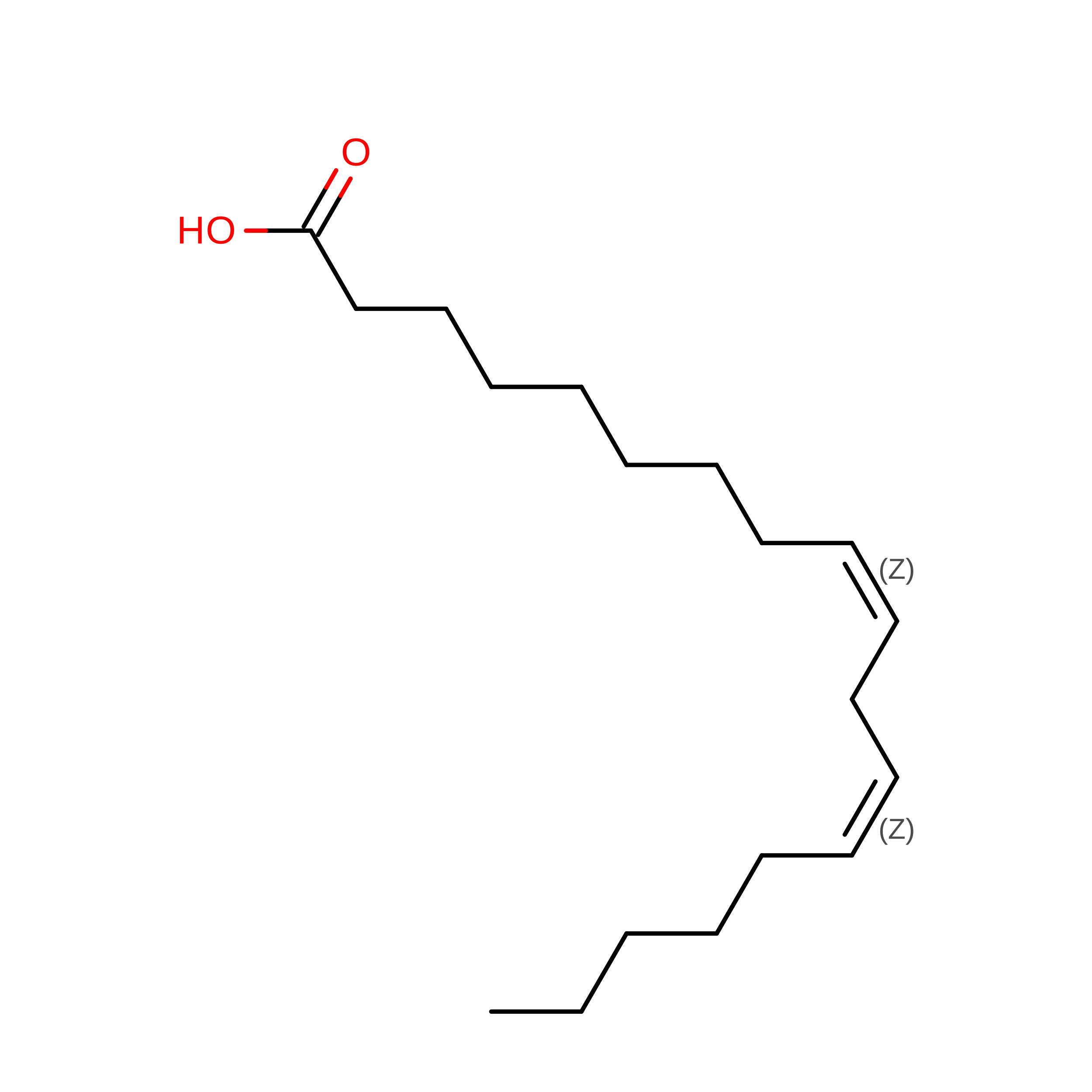
LINOLEIC ACID
C18 H32 O2
OYHQOLUKZRVURQ-HZJYTTRNSA-N
NAG

Query on NAG


Download:Ideal Coordinates CCD File
AA [auth B]
BA [auth B]
DA [auth C]
EA [auth C]
FA [auth C]
AA [auth B],
BA [auth B],
DA [auth C],
EA [auth C],
FA [auth C],
GA [auth C],
H [auth A],
HA [auth C],
I [auth A],
IA [auth C],
J [auth A],
JA [auth C],
K [auth A],
KA [auth C],
L [auth A],
LA [auth C],
M [auth A],
MA [auth C],
N [auth A],
O [auth A],
P [auth A],
Q [auth A],
S [auth B],
T [auth B],
U [auth B],
V [auth B],
W [auth B],
X [auth B],
Y [auth B],
Z [auth B]
2-acetamido-2-deoxy-beta-D-glucopyranose
C8 H15 N O6
OVRNDRQMDRJTHS-FMDGEEDCSA-N

Experimental Data & Validation

Experimental Data

  • Method: ELECTRON MICROSCOPY
  • Resolution: 2.48 Å
  • Aggregation State: PARTICLE 
  • Reconstruction Method: SINGLE PARTICLE 
EM Software:
TaskSoftware PackageVersion
RECONSTRUCTIONRELION
MODEL REFINEMENTPHENIX

Structure Validation

View Full Validation Report


Entry History 

& Funding Information

Deposition Data


Funding OrganizationLocationGrant Number
Biotechnology and Biological Sciences Research Council (BBSRC)United KingdomBB/P000940/1
Wellcome TrustUnited Kingdom202904/Z/16/Z
Wellcome TrustUnited Kingdom206181/Z/17/Z
Biotechnology and Biological Sciences Research Council (BBSRC)United KingdomBB/L01386X/1
Wellcome TrustUnited Kingdom210701/Z/18/Z
Wellcome TrustUnited Kingdom106115/Z/14/Z
Biotechnology and Biological Sciences Research Council (BBSRC)United KingdomBB/R000484/1

Revision History  (Full details and data files)

  • Version 1.0: 2023-02-15
    Type: Initial release
  • Version 1.1: 2024-10-23
    Changes: Data collection, Refinement description, Structure summary