7YM8 | pdb_00007ym8

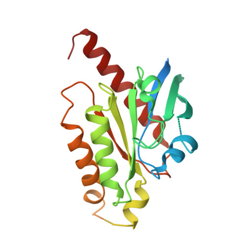
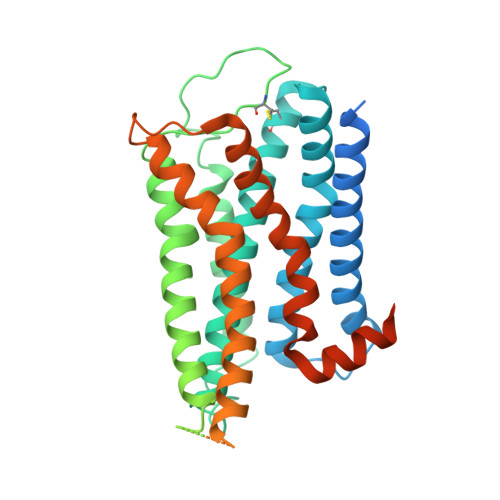
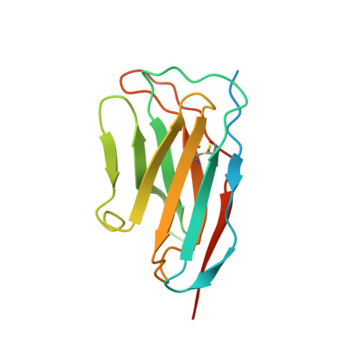
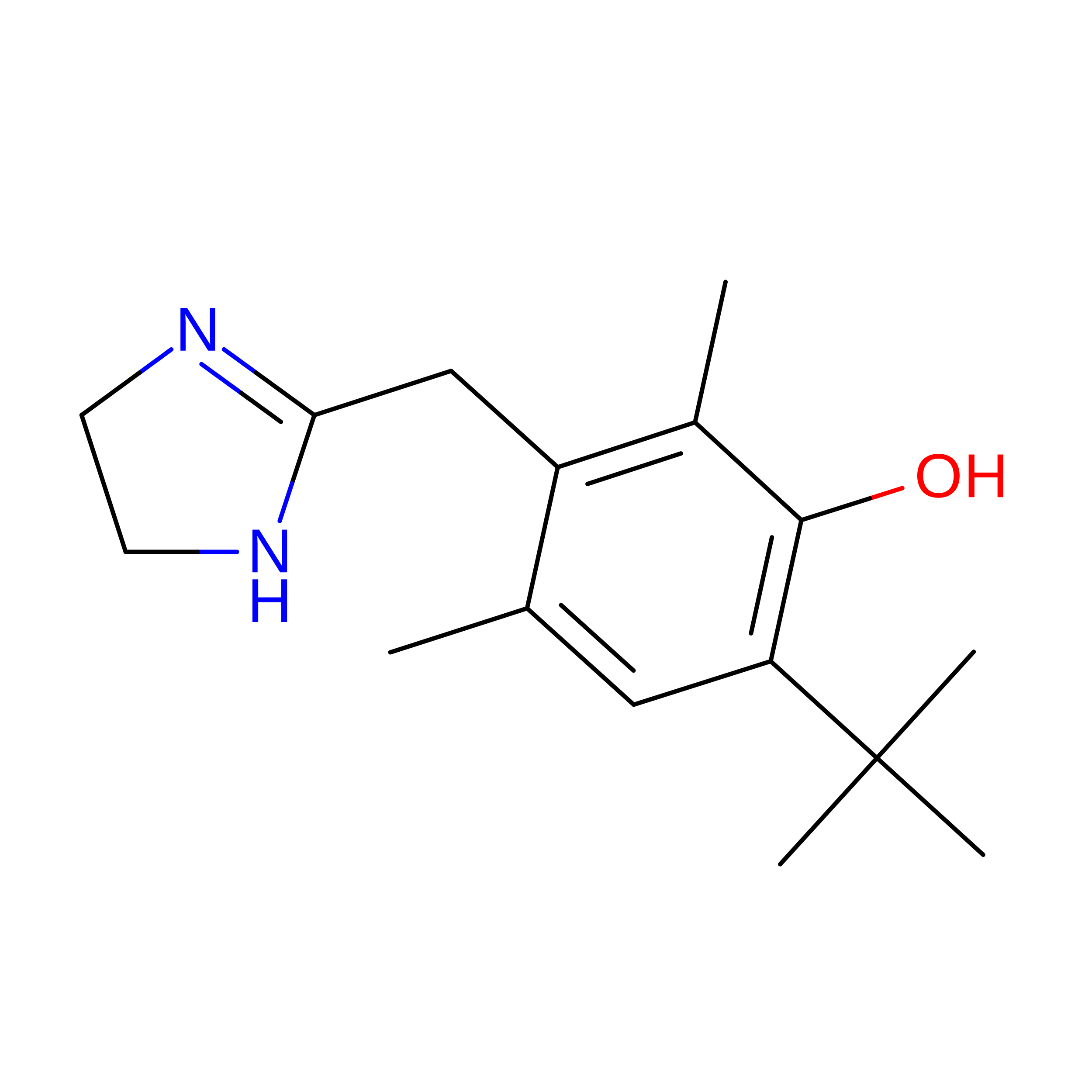
Cryo-EM structure of Nb29-alpha1AAR-miniGsq complex bound to oxymetazoline


Experimental Data Snapshot

  • Method: ELECTRON MICROSCOPY
  • Resolution: 2.92 Å
  • Aggregation State: PARTICLE 
  • Reconstruction Method: SINGLE PARTICLE 

wwPDB Validation 3D Report Full Report

Validation slider image for 7YM8

This is version 1.2 of the entry. See complete history. 

Literature

Structural basis of alpha 1A -adrenergic receptor activation and recognition by an extracellular nanobody.

Toyoda, Y., Zhu, A., Kong, F., Shan, S., Zhao, J., Wang, N., Sun, X., Zhang, L., Yan, C., Kobilka, B.K., Liu, X.

(2023) Nat Commun 14: 3655-3655

  • DOI: https://doi.org/10.1038/s41467-023-39310-x
  • Primary Citation Related Structures: 
    7YM8, 7YMH, 7YMJ

  • PubMed Abstract: 

    The α 1A- adrenergic receptor (α 1A AR) belongs to the family of G protein-coupled receptors that respond to adrenaline and noradrenaline. α 1A AR is involved in smooth muscle contraction and cognitive function. Here, we present three cryo-electron microscopy structures of human α 1A AR bound to the endogenous agonist noradrenaline, its selective agonist oxymetazoline, and the antagonist tamsulosin, with resolutions range from 2.9 Å to 3.5 Å. Our active and inactive α 1A AR structures reveal the activation mechanism and distinct ligand binding modes for noradrenaline compared with other adrenergic receptor subtypes. In addition, we identified a nanobody that preferentially binds to the extracellular vestibule of α 1A AR when bound to the selective agonist oxymetazoline. These results should facilitate the design of more selective therapeutic drugs targeting both orthosteric and allosteric sites in this receptor family.


  • Organizational Affiliation: 
    • School of Medicine, Tsinghua University, Beijing, 100084, China. toyoda.yosuke.2r@kyoto-u.ac.jp.

Macromolecule Content 

  • Total Structure Weight: 98.87 kDa 
  • Atom Count: 4,287 
  • Modeled Residue Count: 556 
  • Deposited Residue Count: 870 
  • Unique protein chains: 3

Macromolecules

Find similar proteins by:|  3D Structure
Entity ID: 1
MoleculeChains  Sequence LengthOrganismDetailsImage
miniGsqA [auth B]246Homo sapiensMutation(s): 0 
Entity Groups
Sequence Clusters30% Identity50% Identity70% Identity90% Identity95% Identity100% Identity
Sequence Annotations
Expand
Reference Sequence
Find similar proteins by:|  3D Structure
Entity ID: 2
MoleculeChains  Sequence LengthOrganismDetailsImage
alpha1A adrenergic receptorB [auth A]480Homo sapiensMutation(s): 0 
EC: 3.2.1.17
Membrane Entity: Yes 
UniProt & NIH Common Fund Data Resources
Find proteins for D9IEF7 (Enterobacteria phage T4)
Explore D9IEF7 
Go to UniProtKB:  D9IEF7
Find proteins for P35348 (Homo sapiens)
Explore P35348 
Go to UniProtKB:  P35348
PHAROS:  P35348
GTEx:  ENSG00000120907 
Entity Groups
Sequence Clusters30% Identity50% Identity70% Identity90% Identity95% Identity100% Identity
UniProt GroupsP35348D9IEF7
Sequence Annotations
Expand
Reference Sequence
Find similar proteins by:|  3D Structure
Entity ID: 3
MoleculeChains  Sequence LengthOrganismDetailsImage
nanobody 29C [auth D]144synthetic constructMutation(s): 0 
Entity Groups
Sequence Clusters30% Identity50% Identity70% Identity90% Identity95% Identity100% Identity
Sequence Annotations
Expand
Reference Sequence

Small Molecules

Binding Affinity Annotations 
IDSourceBinding Affinity
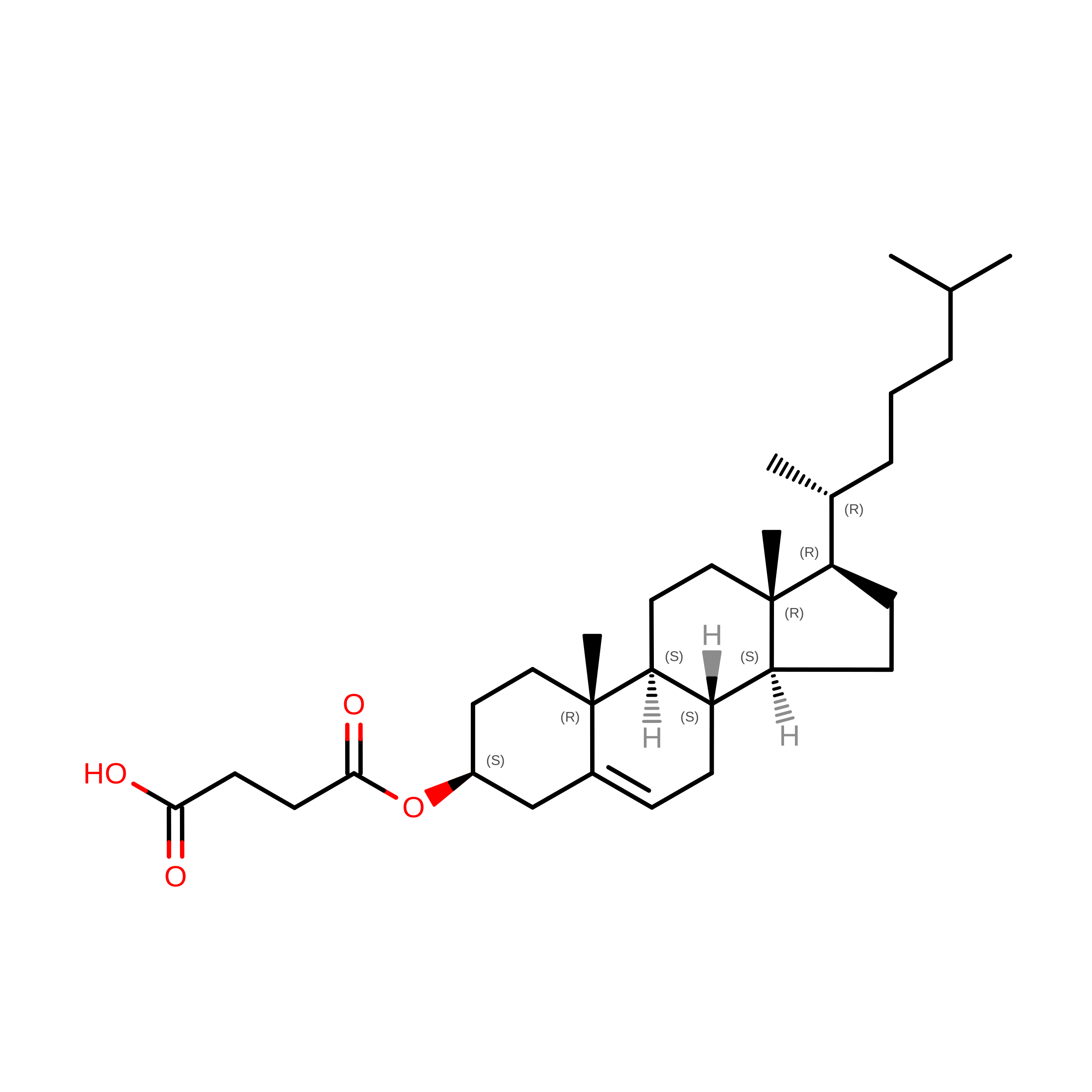
J5C BindingDB:  7YM8 Ki: min: 1.9, max: 2140 (nM) from 12 assay(s)
IC50: 300 (nM) from 1 assay(s)
EC50: min: 20, max: 347 (nM) from 3 assay(s)

Experimental Data & Validation

Experimental Data

  • Method: ELECTRON MICROSCOPY
  • Resolution: 2.92 Å
  • Aggregation State: PARTICLE 
  • Reconstruction Method: SINGLE PARTICLE 
EM Software:
TaskSoftware PackageVersion
MODEL REFINEMENTPHENIX

Structure Validation

View Full Validation Report


Entry History 

& Funding Information

Deposition Data


Funding OrganizationLocationGrant Number
National Natural Science Foundation of China (NSFC)China32122041

Revision History  (Full details and data files)

  • Version 1.0: 2023-07-05
    Type: Initial release
  • Version 1.1: 2024-10-23
    Changes: Data collection, Structure summary
  • Version 1.2: 2025-07-02
    Changes: Data collection