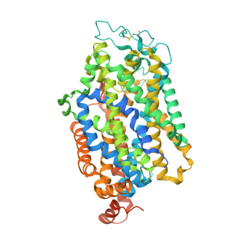
7Y7W

Cryo-EM structure of human GABA transporter GAT1 bound with GABA in NaCl solution in an inward-occluded state at 2.4 angstrom


Experimental Data Snapshot

  • Method: ELECTRON MICROSCOPY
  • Resolution: 2.40 Å
  • Aggregation State: PARTICLE 
  • Reconstruction Method: SINGLE PARTICLE 

wwPDB Validation   3D Report Full Report


This is version 2.1 of the entry. See complete history


Literature

Molecular basis for substrate recognition and transport of human GABA transporter GAT1.

Zhu, A.Huang, J.Kong, F.Tan, J.Lei, J.Yuan, Y.Yan, C.

(2023) Nat Struct Mol Biol 30: 1012-1022

  • DOI: https://doi.org/10.1038/s41594-023-00983-z
  • Primary Citation of Related Structures:  
    7Y7V, 7Y7W, 7Y7Y, 7Y7Z

  • PubMed Abstract: 

    γ-Aminobutyric acid (GABA), an important inhibitory neurotransmitter in the central nervous system, is recycled through specific GABA transporters (GATs). GAT1, which is mainly expressed in the presynaptic terminals of axons, is a potential drug target of neurological disorders due to its essential role in GABA transport. Here we report four cryogenic electron microscopy structures of human GAT1, at resolutions of 2.2-3.2 Å. GAT1 in substrate-free form or in complex with the antiepileptic drug tiagabine exhibits an inward-open conformation. In the presence of GABA or nipecotic acid, inward-occluded structures are captured. The GABA-bound structure reveals an interaction network bridged by hydrogen bonds and ion coordination for GABA recognition. The substrate-free structure unwinds the last helical turn of transmembrane helix TM1a to release sodium ions and substrate. Complemented by structure-guided biochemical analyses, our studies reveal detailed mechanism of GABA recognition and transport, and elucidate mode of action of the inhibitors, nipecotic acid and tiagabine.


  • Organizational Affiliation

    State Key Laboratory of Membrane Biology, Beijing Frontier Research Center for Biological Structure, Beijing Advanced Innovation Center for Structural Biology, Tsinghua-Peking Joint Center for Life Sciences, School of Life Sciences, Tsinghua University, Beijing, China.


Macromolecules
Find similar proteins by:  (by identity cutoff)  |  3D Structure
Entity ID: 1
MoleculeChains Sequence LengthOrganismDetailsImage
Sodium- and chloride-dependent GABA transporter 1632Homo sapiensMutation(s): 0 
Gene Names: SLC6A1GABATRGABT1GAT1
Membrane Entity: Yes 
UniProt & NIH Common Fund Data Resources
Find proteins for P30531 (Homo sapiens)
Explore P30531 
Go to UniProtKB:  P30531
PHAROS:  P30531
GTEx:  ENSG00000157103 
Entity Groups  
Sequence Clusters30% Identity50% Identity70% Identity90% Identity95% Identity100% Identity
UniProt GroupP30531
Glycosylation
Glycosylation Sites: 3Go to GlyGen: P30531-1
Sequence Annotations
Expand
  • Reference Sequence
Small Molecules
Ligands 4 Unique
IDChains Name / Formula / InChI Key2D Diagram3D Interactions
NAG
Query on NAG

Download Ideal Coordinates CCD File 
C [auth A],
D [auth A],
E [auth A]
2-acetamido-2-deoxy-beta-D-glucopyranose
C8 H15 N O6
OVRNDRQMDRJTHS-FMDGEEDCSA-N
ABU (Subject of Investigation/LOI)
Query on ABU

Download Ideal Coordinates CCD File 
B [auth A]GAMMA-AMINO-BUTANOIC ACID
C4 H9 N O2
BTCSSZJGUNDROE-UHFFFAOYSA-N
CL (Subject of Investigation/LOI)
Query on CL

Download Ideal Coordinates CCD File 
H [auth A]CHLORIDE ION
Cl
VEXZGXHMUGYJMC-UHFFFAOYSA-M
NA (Subject of Investigation/LOI)
Query on NA

Download Ideal Coordinates CCD File 
F [auth A],
G [auth A]
SODIUM ION
Na
FKNQFGJONOIPTF-UHFFFAOYSA-N
Experimental Data & Validation

Experimental Data

  • Method: ELECTRON MICROSCOPY
  • Resolution: 2.40 Å
  • Aggregation State: PARTICLE 
  • Reconstruction Method: SINGLE PARTICLE 
EM Software:
TaskSoftware PackageVersion
RECONSTRUCTIONcryoSPARC

Structure Validation

View Full Validation Report


Entry History & Funding Information

Deposition Data


Funding OrganizationLocationGrant Number
Ministry of Science and Technology (MoST, China)China2020YFA0509301

Revision History  (Full details and data files)

  • Version 1.0: 2023-04-26
    Type: Initial release
  • Version 1.1: 2023-08-16
    Changes: Data collection, Database references
  • Version 2.0: 2023-11-15
    Changes: Atomic model, Data collection, Derived calculations
  • Version 2.1: 2024-11-20
    Changes: Data collection, Structure summary