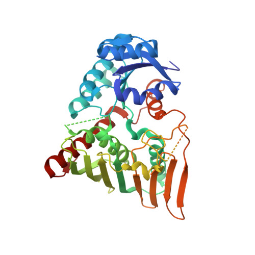
Conformational flexibility enables catalysis of phthalate cis-4,5-dihydrodiol dehydrogenase.
Mahto, J.K., Sharma, M., Neetu, N., Kayastha, A., Aggarwal, S., Kumar, P.(2022) Arch Biochem Biophys 727: 109314-109314
- PubMed: 35667443
- DOI: https://doi.org/10.1016/j.abb.2022.109314
- Primary Citation of Related Structures:
7WZD, 7X1X, 7X2Y - PubMed Abstract:
Phthalate cis-4,5-dihydrodiol dehydrogenase (PhtC), the second enzyme of the phthalate catabolic pathway, catalyzes the dehydrogenation of cis-4,5-dihydrodiol phthalate (DDP). Here, we report the structural and biochemical characterization of PhtC from Comamonas testosteroni KF1 (PhtC KF1 ). With biochemical experiments, we have determined the enzyme's catalytic efficiency (k cat /K m ) with DDP as 2.6 ± 0.5 M -1 s -1 , over 10-fold higher than with cis-3,4-dihydrodiol phthalate (CDP). To understand the structural basis of these reactions, the crystal structures of PhtC KF1 in apo-form, the binary complex with NAD + , and the ternary complex with NAD + and 3-hydroxybenzoate (3HB) were determined. These crystal structures reveal that the binding of 3HB induces a conformational change in the substrate-binding loop. This conformational change causes the opening of the NAD + binding site while trapping the 3HB. The PhtC KF1 crystal structures show that the catalytic domain of PhtC KF1 is larger than that of other structurally characterized homologs and does not align with other cis-diol dehydrogenases. Structural and mutational analysis of the substrate-binding loop residues, Arg164 and Glu167 establish that conformational flexibility of this loop is necessary for positioning the substrate in a catalytically competent pose, as substitution of either of these residues to Ala did not yield the dehydrogenation activity. Further, based on the crystal structures of PhtC KF1 and related structural homologs, a reaction mechanism is proposed. Finally, with the biochemical analysis of a variant M251LPhtC KF1 , the broader substrate specificity of this enzyme is explained.
- Department of Biosciences and Bioengineering, IIT Roorkee, Roorkee, India.
Organizational Affiliation:


