7X2Y | pdb_00007x2y

Crystal Structure of cis-4,5-dihydrodiol phthalate dehydrogenase in complex with NAD+ and 3-Hydroxybenzoate


Experimental Data Snapshot

  • Method: X-RAY DIFFRACTION
  • Resolution: 2.48 Å
  • R-Value Free: 
    0.235 (Depositor), 0.216 (DCC) 
  • R-Value Work: 
    0.192 (Depositor) 
  • R-Value Observed: 
    0.194 (Depositor) 

Starting Model: experimental
View more details

wwPDB Validation 3D Report Full Report

Validation slider image for 7X2Y

Ligand Structure Quality Assessment 


This is version 1.1 of the entry. See complete history

Literature

Conformational flexibility enables catalysis of phthalate cis-4,5-dihydrodiol dehydrogenase.

Mahto, J.K.Sharma, M.Neetu, N.Kayastha, A.Aggarwal, S.Kumar, P.

(2022) Arch Biochem Biophys 727: 109314-109314

  • DOI: https://doi.org/10.1016/j.abb.2022.109314
  • Primary Citation Related Structures: 
    7WZD, 7X1X, 7X2Y

  • PubMed Abstract: 

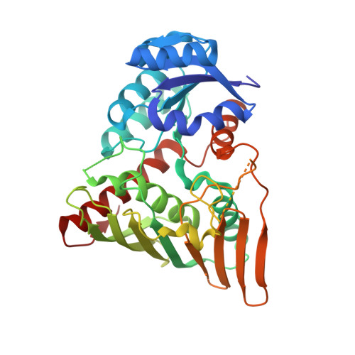
    Phthalate cis-4,5-dihydrodiol dehydrogenase (PhtC), the second enzyme of the phthalate catabolic pathway, catalyzes the dehydrogenation of cis-4,5-dihydrodiol phthalate (DDP). Here, we report the structural and biochemical characterization of PhtC from Comamonas testosteroni KF1 (PhtC KF1 ). With biochemical experiments, we have determined the enzyme's catalytic efficiency (k cat /K m ) with DDP as 2.6 ± 0.5 M -1 s -1 , over 10-fold higher than with cis-3,4-dihydrodiol phthalate (CDP). To understand the structural basis of these reactions, the crystal structures of PhtC KF1 in apo-form, the binary complex with NAD + , and the ternary complex with NAD + and 3-hydroxybenzoate (3HB) were determined. These crystal structures reveal that the binding of 3HB induces a conformational change in the substrate-binding loop. This conformational change causes the opening of the NAD  +  binding site while trapping the 3HB. The PhtC KF1 crystal structures show that the catalytic domain of PhtC KF1 is larger than that of other structurally characterized homologs and does not align with other cis-diol dehydrogenases. Structural and mutational analysis of the substrate-binding loop residues, Arg164 and Glu167 establish that conformational flexibility of this loop is necessary for positioning the substrate in a catalytically competent pose, as substitution of either of these residues to Ala did not yield the dehydrogenation activity. Further, based on the crystal structures of PhtC KF1 and related structural homologs, a reaction mechanism is proposed. Finally, with the biochemical analysis of a variant M251LPhtC KF1 , the broader substrate specificity of this enzyme is explained.


  • Organizational Affiliation
    • Department of Biosciences and Bioengineering, IIT Roorkee, Roorkee, India.

Macromolecule Content 

  • Total Structure Weight: 88.98 kDa 
  • Atom Count: 5,860 
  • Modeled Residue Count: 684 
  • Deposited Residue Count: 784 
  • Unique protein chains: 1

Macromolecules

Find similar proteins by:|  3D Structure
Entity ID: 1
MoleculeChains  Sequence LengthOrganismDetailsImage
4,5-dihydroxyphthalate dehydrogenase
A, B
392Comamonas testosteroni KF-1Mutation(s): 0 
Gene Names: O987_21845
UniProt
Find proteins for A0A076PYH9 (Comamonas testosteroni TK102)
Explore A0A076PYH9 
Go to UniProtKB:  A0A076PYH9
Entity Groups
Sequence Clusters30% Identity50% Identity70% Identity90% Identity95% Identity100% Identity
UniProt GroupA0A076PYH9
Sequence Annotations
Expand
Reference Sequence

Small Molecules

Ligands 5 Unique
IDChains Name / Formula / InChI Key2D Diagram3D Interactions
NAD
(Subject of Investigation/LOI)

Query on NAD


Download:Ideal Coordinates CCD File
I [auth A],
S [auth B]
NICOTINAMIDE-ADENINE-DINUCLEOTIDE
C21 H27 N7 O14 P2
BAWFJGJZGIEFAR-NNYOXOHSSA-N
3HB
(Subject of Investigation/LOI)

Query on 3HB


Download:Ideal Coordinates CCD File
H [auth A],
R [auth B]
3-HYDROXYBENZOIC ACID
C7 H6 O3
IJFXRHURBJZNAO-UHFFFAOYSA-N
PEG

Query on PEG


Download:Ideal Coordinates CCD File
C [auth A]
E [auth A]
L [auth B]
N [auth B]
P [auth B]
C [auth A],
E [auth A],
L [auth B],
N [auth B],
P [auth B],
Q [auth B]
DI(HYDROXYETHYL)ETHER
C4 H10 O3
MTHSVFCYNBDYFN-UHFFFAOYSA-N
PO4

Query on PO4


Download:Ideal Coordinates CCD File
F [auth A],
M [auth B]
PHOSPHATE ION
O4 P
NBIIXXVUZAFLBC-UHFFFAOYSA-K
GOL

Query on GOL


Download:Ideal Coordinates CCD File
D [auth A],
G [auth A],
J [auth A],
K [auth B],
O [auth B]
GLYCEROL
C3 H8 O3
PEDCQBHIVMGVHV-UHFFFAOYSA-N

Experimental Data & Validation

Experimental Data

  • Method: X-RAY DIFFRACTION
  • Resolution: 2.48 Å
  • R-Value Free:  0.235 (Depositor), 0.216 (DCC) 
  • R-Value Work:  0.192 (Depositor) 
  • R-Value Observed: 0.194 (Depositor) 
Space Group: P 21 21 21
Unit Cell:
Length ( Å )Angle ( ˚ )
a = 53.579α = 90
b = 88.315β = 90
c = 162.97γ = 90
Software Package:
Software NamePurpose
REFMACrefinement
PDB_EXTRACTdata extraction
CrysalisProdata reduction
Aimlessdata scaling
MOLREPphasing

Structure Validation

View Full Validation Report


Ligand Structure Quality Assessment 


Entry History 

& Funding Information

Deposition Data


Funding OrganizationLocationGrant Number
Department of Science & Technology (DST, India)IndiaDST/TMD/EWO/WTI/2K19/EWFH/2019/8 (G)

Revision History  (Full details and data files)

  • Version 1.0: 2022-09-14
    Type: Initial release
  • Version 1.1: 2023-11-29
    Changes: Data collection, Refinement description