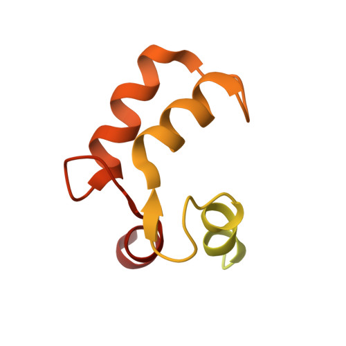
7WZS

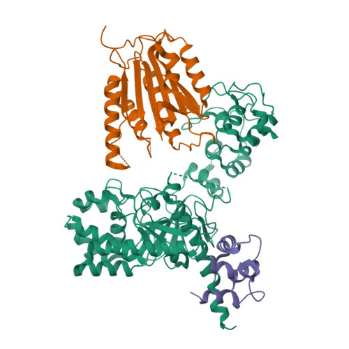
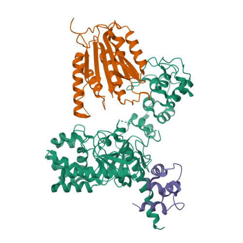
Crystal structure of Chromobacterium violaceum effector CopC in complex with host calmodulin and caspase-7


Experimental Data Snapshot

  • Method: X-RAY DIFFRACTION
  • Resolution: 3.60 Å
  • R-Value Free: 0.314 
  • R-Value Work: 0.274 
  • R-Value Observed: 0.278 

Starting Model: experimental
View more details

wwPDB Validation   3D Report Full Report


This is version 1.2 of the entry. See complete history


Literature

Calmodulin Binding Activates Chromobacterium CopC Effector to ADP-Riboxanate Host Apoptotic Caspases.

Liu, Y.Zeng, H.Hou, Y.Li, Z.Li, L.Song, X.Ding, J.Shao, F.Xu, Y.

(2022) mBio 13: e0069022-e0069022

  • DOI: https://doi.org/10.1128/mbio.00690-22
  • Primary Citation of Related Structures:  
    7WZS

  • PubMed Abstract: 

    Blocking host cell death is an important virulence strategy employed by many bacterial pathogens. We recently reported that Shigella flexneri inhibits host pyroptosis by delivering a type III secretion system (T3SS) effector OspC3 that catalyzes a novel arginine ADP-riboxanation modification on caspase-4/11. Here, we investigated the OspC3 homologue CopC from Chromobacterium violaceum, an opportunistic but sometimes deadly bacterial pathogen. CopC bears the same arginine ADP-riboxanase activity as OspC3, but with a different substrate specificity. Through proteomic analysis, we first identified host calmodulin (CaM) as a binding partner of CopC. The analyses additionally revealed that CopC preferably modifies apoptotic caspases including caspase-7, -8 and -9. This results in suppression of both extrinsic and intrinsic apoptosis programs in C. violaceum-infected cells. Biochemical reconstitution showed that CopC requires binding to CaM, specifically in the calcium-free state, to achieve efficient ADP-riboxanation of the caspases. We determined crystal structure of the CaM-CopC-CASP7 ternary complex, which illustrates the caspase recognition mechanism and a unique CaM-binding mode in CopC. Structure-directed mutagenesis validated the functional significance of CaM binding for stimulating CopC modification of its caspase substrates. CopC adopts an ADP-ribosyltransferase-like fold with a unique His-Phe-Glu catalytic triad, featuring two acidic residues critical for site-specific arginine ADP-riboxanation. Our study expands and deepens our understanding of the OspC family of ADP-riboxanase effectors. IMPORTANCE Programmed cell death is a suicidal defense mechanism for eukaryotes to combat pathogen infection. In the evolutionary arms race with the host, bacteria are endowed with ingenious tactics to block host cell death to facilitate their replication. Here, we report that the C. violaceum effector CopC ADP-riboxanates caspase-7/8/9, enabled by interacting with the host factor calmodulin, to block host cell apoptosis, illustrating a unique and sophisticated strategy adopted by the pathogen to counteract host defense.


  • Organizational Affiliation

    Graduate Program in Chinese Academy of Medical Sciences and Peking Union Medical College, Beijing, China.


Macromolecules
Find similar proteins by:  (by identity cutoff)  |  3D Structure
Entity ID: 1
MoleculeChains Sequence LengthOrganismDetailsImage
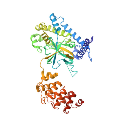
CopC443Chromobacterium violaceum ATCC 12472Mutation(s): 0 
Gene Names: CV_2038
EC: 4.3.99
UniProt
Find proteins for Q7NWF2 (Chromobacterium violaceum (strain ATCC 12472 / DSM 30191 / JCM 1249 / CCUG 213 / NBRC 12614 / NCIMB 9131 / NCTC 9757 / MK))
Explore Q7NWF2 
Go to UniProtKB:  Q7NWF2
Entity Groups  
Sequence Clusters30% Identity50% Identity70% Identity90% Identity95% Identity100% Identity
UniProt GroupQ7NWF2
Sequence Annotations
Expand
  • Reference Sequence
Find similar proteins by:  (by identity cutoff)  |  3D Structure
Entity ID: 2
MoleculeChains Sequence LengthOrganismDetailsImage
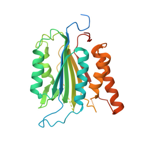
Caspase-7289Homo sapiensMutation(s): 1 
Gene Names: CASP7
EC: 3.4.22.60
UniProt & NIH Common Fund Data Resources
Find proteins for P55210 (Homo sapiens)
Explore P55210 
Go to UniProtKB:  P55210
PHAROS:  P55210
GTEx:  ENSG00000165806 
Entity Groups  
Sequence Clusters30% Identity50% Identity70% Identity90% Identity95% Identity100% Identity
UniProt GroupP55210
Sequence Annotations
Expand
  • Reference Sequence
Find similar proteins by:  (by identity cutoff)  |  3D Structure
Entity ID: 3
MoleculeChains Sequence LengthOrganismDetailsImage
Calmodulin-1153Homo sapiensMutation(s): 0 
Gene Names: CALM1CALMCAMCAM1
UniProt & NIH Common Fund Data Resources
Find proteins for P0DP23 (Homo sapiens)
Explore P0DP23 
Go to UniProtKB:  P0DP23
PHAROS:  P0DP23
GTEx:  ENSG00000198668 
Entity Groups  
Sequence Clusters30% Identity50% Identity70% Identity90% Identity95% Identity100% Identity
UniProt GroupP0DP23
Sequence Annotations
Expand
  • Reference Sequence
Experimental Data & Validation

Experimental Data

  • Method: X-RAY DIFFRACTION
  • Resolution: 3.60 Å
  • R-Value Free: 0.314 
  • R-Value Work: 0.274 
  • R-Value Observed: 0.278 
  • Space Group: P 43 21 2
Unit Cell:
Length ( Å )Angle ( ˚ )
a = 90.871α = 90
b = 90.871β = 90
c = 426.106γ = 90
Software Package:
Software NamePurpose
PHENIXrefinement
XDSdata reduction
XSCALEdata scaling
PHASERphasing

Structure Validation

View Full Validation Report


Entry History & Funding Information

Deposition Data

  • Released Date: 2022-05-18 
  • Deposition Author(s): Ding, J.

Funding OrganizationLocationGrant Number
Chinese Academy of SciencesChinaXDB29020202

Revision History  (Full details and data files)

  • Version 1.0: 2022-05-18
    Type: Initial release
  • Version 1.1: 2022-07-13
    Changes: Database references
  • Version 1.2: 2023-11-29
    Changes: Data collection, Refinement description