7V9B | pdb_00007v9b

Crystal Structure of 14-3-3 epsilon with FOXO3a peptide


Experimental Data Snapshot

  • Method: X-RAY DIFFRACTION
  • Resolution: 1.85 Å
  • R-Value Free: 
    0.189 (Depositor), 0.193 (DCC) 
  • R-Value Work: 
    0.167 (Depositor), 0.171 (DCC) 
  • R-Value Observed: 
    0.168 (Depositor) 

Starting Model: experimental
View more details

wwPDB Validation 3D Report Full Report

Validation slider image for 7V9B

Ligand Structure Quality Assessment 


This is version 1.2 of the entry. See complete history

Literature

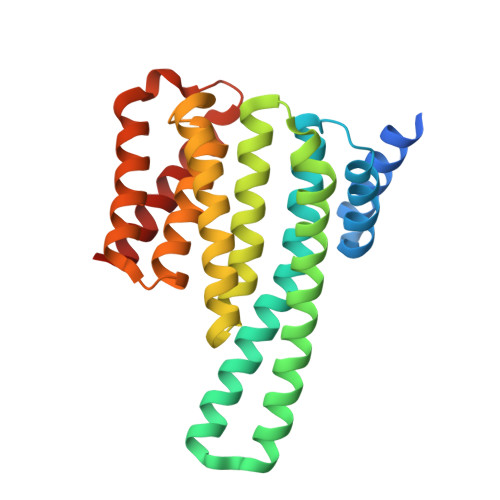
Structure of a 14-3-3 epsilon :FOXO3a pS253 Phosphopeptide Complex Reveals 14-3-3 Isoform-Specific Binding of Forkhead Box Class O Transcription Factor (FOXO) Phosphoproteins.

Mathivanan, S.Chunchagatta Lakshman, P.K.Singh, M.Giridharan, S.Sathish, K.Hurakadli, M.A.Bharatham, K.Kamariah, N.

(2022) ACS Omega 7: 24344-24352

  • DOI: https://doi.org/10.1021/acsomega.2c01700
  • Primary Citation Related Structures: 
    7V9B

  • PubMed Abstract: 

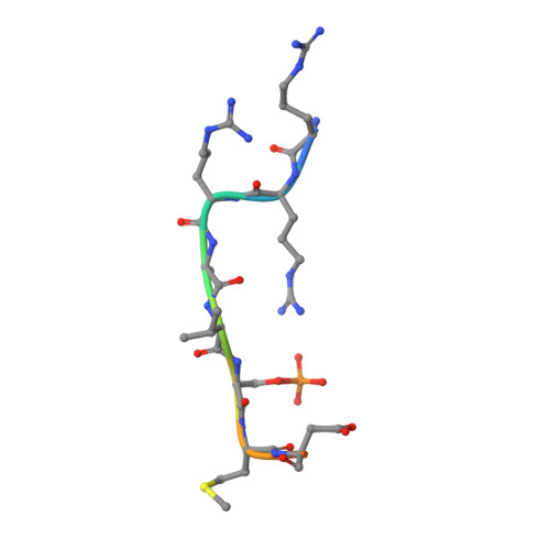
    The transcriptional activity of Forkhead Box O3 (FOXO3a) is inactivated by AKT-mediated phosphorylation on Serine 253 (S253), which enables FOXO3a binding to 14-3-3. Phosphorylated FOXO3a binding to 14-3-3 facilitates the nuclear exclusion of FOXO3a, causing cancer cell proliferation. The FOXO3a/14-3-3 interaction has, therefore, emerged as an important therapeutic target. Here, we report a comprehensive analysis using fluorescence polarization, isothermal titration calorimetry, small-angle X-ray scattering, X-ray crystallography, and molecular dynamics simulations to gain molecular-level insights into the interaction of FOXO3a pS253 phosphopeptide with 14-3-3ε. A high-resolution structure of the fluorophore-labeled FOXO3a pS253 :14-3-3ε complex revealed a distinct mode of interaction compared to other 14-3-3 phosphopeptide complexes. FOXO3a pS253 phosphopeptide showed significant structural difference in the positions of the -3 and -4 Arg residues relative to pSer, compared to that of a similar phosphopeptide, FOXO1 pS256 bound to 14-3-3σ. Moreover, molecular dynamics studies show that the significant structural changes and molecular interactions noticed in the crystal structure of FOXO3a pS253 :14-3-3ε are preserved over the course of the simulation. Thus, this study reveals structural differences between the binding to 14-3-3 isoforms of FOXO1 pS256 versus FOXO3a pS253 , providing a framework for the rational design of isoform-specific FOXO/14-3-3 protein-protein interaction inhibitors for therapy.


  • Organizational Affiliation
    • Centre for Chemical Biology & Therapeutics, inStem & NCBS, Bellary Road, Bangalore 560065, India.

Macromolecule Content 

  • Total Structure Weight: 30.46 kDa 
  • Atom Count: 2,148 
  • Modeled Residue Count: 239 
  • Deposited Residue Count: 260 
  • Unique protein chains: 2

Macromolecules

Find similar proteins by:|  3D Structure
Entity ID: 1
MoleculeChains  Sequence LengthOrganismDetailsImage
YWHAE/FAM22B fusion protein249Homo sapiensMutation(s): 0 
Gene Names: YWHAE/FAM22B fusion
UniProt & NIH Common Fund Data Resources
Find proteins for P62258 (Homo sapiens)
Explore P62258 
Go to UniProtKB:  P62258
PHAROS:  P62258
GTEx:  ENSG00000108953 
Entity Groups
Sequence Clusters30% Identity50% Identity70% Identity90% Identity95% Identity100% Identity
UniProt GroupP62258
Sequence Annotations
Expand
Reference Sequence
Find similar proteins by:  Sequence   |   3D Structure  
Entity ID: 2
MoleculeChains  Sequence LengthOrganismDetailsImage
ARG-ARG-ARG-ALA-VAL-SEP-MET-ASP-ASN-SER-ASN11Homo sapiensMutation(s): 0 
UniProt & NIH Common Fund Data Resources
Find proteins for O43524 (Homo sapiens)
Explore O43524 
Go to UniProtKB:  O43524
PHAROS:  O43524
GTEx:  ENSG00000118689 
Entity Groups
Sequence Clusters30% Identity50% Identity70% Identity90% Identity95% Identity100% Identity
UniProt GroupO43524
Sequence Annotations
Expand
Reference Sequence

Small Molecules

Ligands 4 Unique
IDChains Name / Formula / InChI Key2D Diagram3D Interactions
323
(Subject of Investigation/LOI)

Query on 323


Download:Ideal Coordinates CCD File
G [auth B]2-[3,6-bis(dimethylamino)xanthen-9-yl]-5-methanoyl-benzoate
C25 H22 N2 O4
ZFGLYCCNMJCDCP-UHFFFAOYSA-N
MPD

Query on MPD


Download:Ideal Coordinates CCD File
E [auth A](4S)-2-METHYL-2,4-PENTANEDIOL
C6 H14 O2
SVTBMSDMJJWYQN-YFKPBYRVSA-N
GOL

Query on GOL


Download:Ideal Coordinates CCD File
C [auth A],
D [auth A]
GLYCEROL
C3 H8 O3
PEDCQBHIVMGVHV-UHFFFAOYSA-N
IMD

Query on IMD


Download:Ideal Coordinates CCD File
F [auth A]IMIDAZOLE
C3 H5 N2
RAXXELZNTBOGNW-UHFFFAOYSA-O
Modified Residues  1 Unique
IDChains TypeFormula2D DiagramParent
SEP
Query on SEP
B
L-PEPTIDE LINKINGC3 H8 N O6 PSER

Experimental Data & Validation

Experimental Data

  • Method: X-RAY DIFFRACTION
  • Resolution: 1.85 Å
  • R-Value Free:  0.189 (Depositor), 0.193 (DCC) 
  • R-Value Work:  0.167 (Depositor), 0.171 (DCC) 
  • R-Value Observed: 0.168 (Depositor) 
Space Group: C 2 2 21
Unit Cell:
Length ( Å )Angle ( ˚ )
a = 77.88α = 90
b = 81.04β = 90
c = 83.28γ = 90
Software Package:
Software NamePurpose
REFMACrefinement
iMOSFLMdata reduction
SCALAdata scaling
PHASERphasing

Structure Validation

View Full Validation Report


Ligand Structure Quality Assessment 


Entry History 

& Funding Information

Deposition Data


Funding OrganizationLocationGrant Number
Department of Biotechnology (DBT, India)India--

Revision History  (Full details and data files)

  • Version 1.0: 2022-08-31
    Type: Initial release
  • Version 1.1: 2022-09-07
    Changes: Derived calculations
  • Version 1.2: 2023-11-29
    Changes: Data collection, Refinement description