7V9B

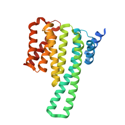
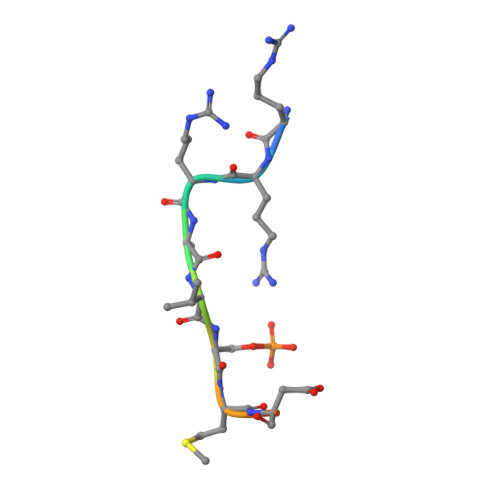
Crystal Structure of 14-3-3 epsilon with FOXO3a peptide


Experimental Data Snapshot

  • Method: X-RAY DIFFRACTION
  • Resolution: 1.85 Å
  • R-Value Free: 0.189 
  • R-Value Work: 0.167 
  • R-Value Observed: 0.168 

Starting Model: experimental
View more details

wwPDB Validation   3D Report Full Report


Ligand Structure Quality Assessment 


This is version 1.2 of the entry. See complete history


Literature

Structure of a 14-3-3 epsilon :FOXO3a pS253 Phosphopeptide Complex Reveals 14-3-3 Isoform-Specific Binding of Forkhead Box Class O Transcription Factor (FOXO) Phosphoproteins.

Mathivanan, S.Chunchagatta Lakshman, P.K.Singh, M.Giridharan, S.Sathish, K.Hurakadli, M.A.Bharatham, K.Kamariah, N.

(2022) ACS Omega 7: 24344-24352

  • DOI: https://doi.org/10.1021/acsomega.2c01700
  • Primary Citation of Related Structures:  
    7V9B

  • PubMed Abstract: 

    The transcriptional activity of Forkhead Box O3 (FOXO3a) is inactivated by AKT-mediated phosphorylation on Serine 253 (S253), which enables FOXO3a binding to 14-3-3. Phosphorylated FOXO3a binding to 14-3-3 facilitates the nuclear exclusion of FOXO3a, causing cancer cell proliferation. The FOXO3a/14-3-3 interaction has, therefore, emerged as an important therapeutic target. Here, we report a comprehensive analysis using fluorescence polarization, isothermal titration calorimetry, small-angle X-ray scattering, X-ray crystallography, and molecular dynamics simulations to gain molecular-level insights into the interaction of FOXO3a pS253 phosphopeptide with 14-3-3ε. A high-resolution structure of the fluorophore-labeled FOXO3a pS253 :14-3-3ε complex revealed a distinct mode of interaction compared to other 14-3-3 phosphopeptide complexes. FOXO3a pS253 phosphopeptide showed significant structural difference in the positions of the -3 and -4 Arg residues relative to pSer, compared to that of a similar phosphopeptide, FOXO1 pS256 bound to 14-3-3σ. Moreover, molecular dynamics studies show that the significant structural changes and molecular interactions noticed in the crystal structure of FOXO3a pS253 :14-3-3ε are preserved over the course of the simulation. Thus, this study reveals structural differences between the binding to 14-3-3 isoforms of FOXO1 pS256 versus FOXO3a pS253 , providing a framework for the rational design of isoform-specific FOXO/14-3-3 protein-protein interaction inhibitors for therapy.


  • Organizational Affiliation

    Centre for Chemical Biology & Therapeutics, inStem & NCBS, Bellary Road, Bangalore 560065, India.


Macromolecules
Find similar proteins by:  (by identity cutoff)  |  3D Structure
Entity ID: 1
MoleculeChains Sequence LengthOrganismDetailsImage
YWHAE/FAM22B fusion protein249Homo sapiensMutation(s): 0 
Gene Names: YWHAE/FAM22B fusion
UniProt & NIH Common Fund Data Resources
Find proteins for P62258 (Homo sapiens)
Explore P62258 
Go to UniProtKB:  P62258
PHAROS:  P62258
GTEx:  ENSG00000108953 
Entity Groups  
Sequence Clusters30% Identity50% Identity70% Identity90% Identity95% Identity100% Identity
UniProt GroupP62258
Sequence Annotations
Expand
  • Reference Sequence

Find similar proteins by:  Sequence   |   3D Structure  

Entity ID: 2
MoleculeChains Sequence LengthOrganismDetailsImage
ARG-ARG-ARG-ALA-VAL-SEP-MET-ASP-ASN-SER-ASN11Homo sapiensMutation(s): 0 
UniProt & NIH Common Fund Data Resources
Find proteins for O43524 (Homo sapiens)
Explore O43524 
Go to UniProtKB:  O43524
PHAROS:  O43524
GTEx:  ENSG00000118689 
Entity Groups  
Sequence Clusters30% Identity50% Identity70% Identity90% Identity95% Identity100% Identity
UniProt GroupO43524
Sequence Annotations
Expand
  • Reference Sequence
Small Molecules
Modified Residues  1 Unique
IDChains TypeFormula2D DiagramParent
SEP
Query on SEP
B
L-PEPTIDE LINKINGC3 H8 N O6 PSER
Experimental Data & Validation

Experimental Data

  • Method: X-RAY DIFFRACTION
  • Resolution: 1.85 Å
  • R-Value Free: 0.189 
  • R-Value Work: 0.167 
  • R-Value Observed: 0.168 
  • Space Group: C 2 2 21
Unit Cell:
Length ( Å )Angle ( ˚ )
a = 77.88α = 90
b = 81.04β = 90
c = 83.28γ = 90
Software Package:
Software NamePurpose
REFMACrefinement
iMOSFLMdata reduction
SCALAdata scaling
PHASERphasing

Structure Validation

View Full Validation Report


Ligand Structure Quality Assessment 


Entry History & Funding Information

Deposition Data


Funding OrganizationLocationGrant Number
Department of Biotechnology (DBT, India)India--

Revision History  (Full details and data files)

  • Version 1.0: 2022-08-31
    Type: Initial release
  • Version 1.1: 2022-09-07
    Changes: Derived calculations
  • Version 1.2: 2023-11-29
    Changes: Data collection, Refinement description