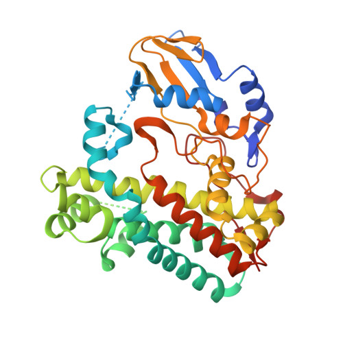
The Cytochrome P450 OxyA from the Kistamicin Biosynthesis Cyclization Cascade is Highly Sensitive to Oxidative Damage.
Greule, A., Izore, T., Machell, D., Hansen, M.H., Schoppet, M., De Voss, J.J., Charkoudian, L.K., Schittenhelm, R.B., Harmer, J.R., Cryle, M.J.(2022) Front Chem 10: 868240-868240
- PubMed: 35464232 Search on PubMedSearch on PubMed Central
- DOI: https://doi.org/10.3389/fchem.2022.868240
- Primary Citation Related Structures:
7TTA, 7TTB, 7TTO, 7TTP, 7TTQ - PubMed Abstract:
Cytochrome P450 enzymes (P450s) are a superfamily of monooxygenases that utilize a cysteine thiolate-ligated heme moiety to perform a wide range of demanding oxidative transformations. Given the oxidative power of the active intermediate formed within P450s during their active cycle, it is remarkable that these enzymes can avoid auto-oxidation and retain the axial cysteine ligand in the deprotonated-and thus highly acidic-thiolate form. While little is known about the process of heme incorporation during P450 folding, there is an overwhelming preference for one heme orientation within the P450 active site. Indeed, very few structures to date contain an alternate heme orientation, of which two are OxyA homologs from glycopeptide antibiotic (GPA) biosynthesis. Given the apparent preference for the unusual heme orientation shown by OxyA enzymes, we investigated the OxyA homolog from kistamicin biosynthesis (OxyA kis ), which is an atypical GPA. We determined that OxyA kis is highly sensitive to oxidative damage by peroxide, with both UV and EPR measurements showing rapid bleaching of the heme signal. We determined the structure of OxyA kis and found a mixed population of heme orientations present in this enzyme. Our analysis further revealed the possible modification of the heme moiety, which was only present in samples where the alternate heme orientation was present in the protein. These results suggest that the typical heme orientation in cytochrome P450s can help prevent potential damage to the heme-and hence deactivation of the enzyme-during P450 catalysis. It also suggests that some P450 enzymes involved in GPA biosynthesis may be especially prone to oxidative damage due to the heme orientation found in their active sites.
- Department of Biochemistry and Molecular Biology, The Monash Biomedicine Discovery Institute, Monash University, Clayton, VIC, Australia.
Organizational Affiliation: