7RSS | pdb_00007rss

Kod-RI incorporating DNA, n+2


Experimental Data Snapshot

  • Method: X-RAY DIFFRACTION
  • Resolution: 2.71 Å
  • R-Value Free: 
    0.225 (Depositor), 0.224 (DCC) 
  • R-Value Work: 
    0.159 (Depositor), 0.160 (DCC) 
  • R-Value Observed: 
    0.164 (Depositor) 

Starting Model: experimental
View more details

wwPDB Validation 3D Report Full Report

Validation slider image for 7RSS

This is version 1.3 of the entry. See complete history

Literature

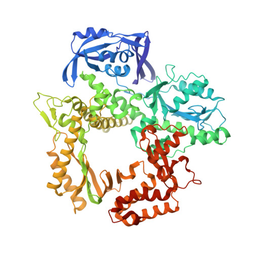
Crystallographic analysis of engineered polymerases synthesizing phosphonomethylthreosyl nucleic acid.

Hajjar, M.Chim, N.Liu, C.Herdewijn, P.Chaput, J.C.

(2022) Nucleic Acids Res 50: 9663-9674

  • DOI: https://doi.org/10.1093/nar/gkac792
  • Primary Citation Related Structures: 
    7RSR, 7RSS, 7TQW

  • PubMed Abstract: 

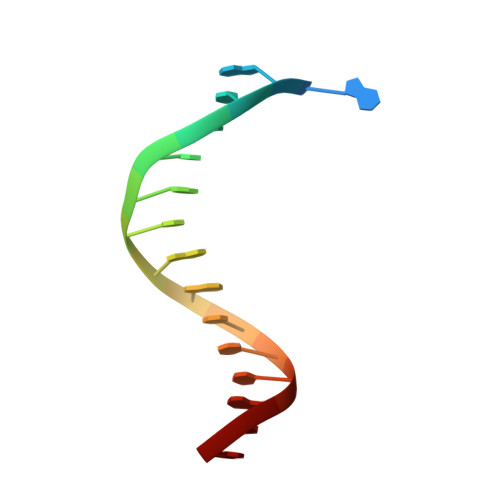
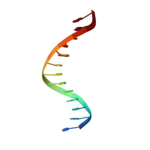
    Xeno-nucleic acids (XNAs) are synthetic genetic polymers with backbone structures composed of non-ribose or non-deoxyribose sugars. Phosphonomethylthreosyl nucleic acid (pTNA), a type of XNA that does not base pair with DNA or RNA, has been suggested as a possible genetic material for storing synthetic biology information in cells. A critical step in this process is the synthesis of XNA episomes using laboratory-evolved polymerases to copy DNA information into XNA. Here, we investigate the polymerase recognition of pTNA nucleotides using X-ray crystallography to capture the post-catalytic complex of engineered polymerases following the sequential addition of two pTNA nucleotides onto the 3'-end of a DNA primer. High-resolution crystal structures reveal that the polymerase mediates Watson-Crick base pairing between the extended pTNA adducts and the DNA template. Comparative analysis studies demonstrate that the sugar conformation and backbone position of pTNA are structurally more similar to threose nucleic acid than DNA even though pTNA and DNA share the same six-atom backbone repeat length. Collectively, these findings provide new insight into the structural determinants that guide the enzymatic synthesis of an orthogonal genetic polymer, and may lead to the discovery of new variants that function with enhanced activity.


  • Organizational Affiliation
    • Department of Pharmaceutical Sciences, University of California, Irvine, CA 92697-3958, USA.

Macromolecule Content 

  • Total Structure Weight: 99.01 kDa 
  • Atom Count: 6,883 
  • Modeled Residue Count: 784 
  • Deposited Residue Count: 803 
  • Unique protein chains: 1
  • Unique nucleic acid chains: 2

Macromolecules


Find similar proteins by:|  3D Structure
Entity ID: 1
MoleculeChains  Sequence LengthOrganismDetailsImage
DNA polymerase774Thermococcus kodakarensisMutation(s): 0 
EC: 2.7.7.7
UniProt
Find proteins for D0VWU9 (Thermococcus kodakarensis (strain ATCC BAA-918 / JCM 12380 / KOD1))
Explore D0VWU9 
Go to UniProtKB:  D0VWU9
Entity Groups
Sequence Clusters30% Identity50% Identity70% Identity90% Identity95% Identity100% Identity
UniProt GroupD0VWU9
Sequence Annotations
Expand
Reference Sequence
Find similar nucleic acids by:  Sequence
Entity ID: 2
MoleculeChains LengthOrganismImage
TemplateB [auth T]16synthetic construct
Sequence Annotations
Expand
Reference Sequence
Find similar nucleic acids by:  Sequence
Entity ID: 3
MoleculeChains LengthOrganismImage
PrimerC [auth P]13synthetic construct
Sequence Annotations
Expand
Reference Sequence

Experimental Data & Validation

Experimental Data

  • Method: X-RAY DIFFRACTION
  • Resolution: 2.71 Å
  • R-Value Free:  0.225 (Depositor), 0.224 (DCC) 
  • R-Value Work:  0.159 (Depositor), 0.160 (DCC) 
  • R-Value Observed: 0.164 (Depositor) 
Space Group: P 2 21 21
Unit Cell:
Length ( Å )Angle ( ˚ )
a = 66.259α = 90
b = 112β = 90
c = 149.197γ = 90
Software Package:
Software NamePurpose
MOSFLMdata reduction
Aimlessdata scaling
PHASERphasing
PHENIXrefinement

Structure Validation

View Full Validation Report


Entry History 

& Funding Information

Deposition Data


Funding OrganizationLocationGrant Number
National Science Foundation (NSF, United States)United States--

Revision History  (Full details and data files)

  • Version 1.0: 2022-08-24
    Type: Initial release
  • Version 1.1: 2022-09-28
    Changes: Database references
  • Version 1.2: 2022-10-05
    Changes: Database references
  • Version 1.3: 2023-10-18
    Changes: Data collection, Refinement description