7RNW | pdb_00007rnw

SARS-CoV-2 Main Protease in complex with a cyclic peptide inhibitor


Experimental Data Snapshot

  • Method: X-RAY DIFFRACTION
  • Resolution: 2.35 Å
  • R-Value Free: 
    0.231 (Depositor), 0.214 (DCC) 
  • R-Value Work: 
    0.171 (Depositor) 
  • R-Value Observed: 
    0.174 (Depositor) 

Starting Model: experimental
View more details

wwPDB Validation 3D Report Full Report

Validation slider image for 7RNW

This is version 1.2 of the entry. See complete history

Literature

Antiviral cyclic peptides targeting the main protease of SARS-CoV-2.

Johansen-Leete, J.Ullrich, S.Fry, S.E.Frkic, R.Bedding, M.J.Aggarwal, A.Ashhurst, A.S.Ekanayake, K.B.Mahawaththa, M.C.Sasi, V.M.Luedtke, S.Ford, D.J.O'Donoghue, A.J.Passioura, T.Larance, M.Otting, G.Turville, S.Jackson, C.J.Nitsche, C.Payne, R.J.

(2022) Chem Sci 13: 3826-3836

  • DOI: https://doi.org/10.1039/d1sc06750h
  • Primary Citation Related Structures: 
    7RNW

  • PubMed Abstract: 

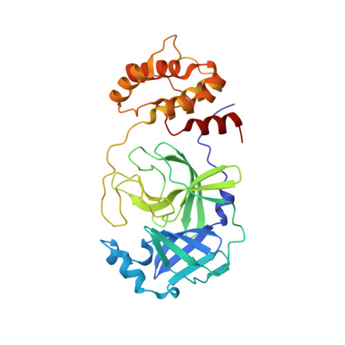
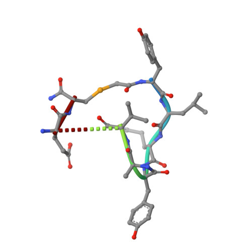
    Antivirals that specifically target SARS-CoV-2 are needed to control the COVID-19 pandemic. The main protease (M pro ) is essential for SARS-CoV-2 replication and is an attractive target for antiviral development. Here we report the use of the Random nonstandard Peptide Integrated Discovery (RaPID) mRNA display on a chemically cross-linked SARS-CoV-2 M pro dimer, which yielded several high-affinity thioether-linked cyclic peptide inhibitors of the protease. Structural analysis of M pro complexed with a selenoether analogue of the highest-affinity peptide revealed key binding interactions, including glutamine and leucine residues in sites S1 and S2, respectively, and a binding epitope straddling both protein chains in the physiological dimer. Several of these M pro peptide inhibitors possessed antiviral activity against SARS-CoV-2 in vitro with EC 50 values in the low micromolar range. These cyclic peptides serve as a foundation for the development of much needed antivirals that specifically target SARS-CoV-2.


  • Organizational Affiliation
    • School of Chemistry, The University of Sydney Sydney NSW 2006 Australia richard.payne@sydney.edu.au.

Macromolecule Content 

  • Total Structure Weight: 142.95 kDa 
  • Atom Count: 9,819 
  • Modeled Residue Count: 1,259 
  • Deposited Residue Count: 1,284 
  • Unique protein chains: 2

Macromolecules

Find similar proteins by:|  3D Structure
Entity ID: 1
MoleculeChains  Sequence LengthOrganismDetailsImage
3C-like proteinase
A, B, C, D
306Severe acute respiratory syndrome coronavirus 2Mutation(s): 0 
Gene Names: rep1a-1b
EC: 3.4.22.69
UniProt
Find proteins for P0DTD1 (Severe acute respiratory syndrome coronavirus 2)
Explore P0DTD1 
Go to UniProtKB:  P0DTD1
Entity Groups
Sequence Clusters30% Identity50% Identity70% Identity90% Identity95% Identity100% Identity
UniProt GroupP0DTD1
Sequence Annotations
Expand
Reference Sequence
Find similar proteins by:  Sequence   |   3D Structure  
Entity ID: 2
MoleculeChains  Sequence LengthOrganismDetailsImage
ACE-DTY-LEU-GLN-TYR-ALA-VAL-LEU-ARG-HIS-LYS-ARG-ARG-GLU-SECE [auth X],
F [auth Y],
G [auth Z],
H [auth W]
15synthetic constructMutation(s): 0 

Experimental Data & Validation

Experimental Data

  • Method: X-RAY DIFFRACTION
  • Resolution: 2.35 Å
  • R-Value Free:  0.231 (Depositor), 0.214 (DCC) 
  • R-Value Work:  0.171 (Depositor) 
  • R-Value Observed: 0.174 (Depositor) 
Space Group: P 1 21 1
Unit Cell:
Length ( Å )Angle ( ˚ )
a = 48.36α = 90
b = 201.726β = 113.49
c = 60.543γ = 90
Software Package:
Software NamePurpose
REFMACrefinement
Aimlessdata scaling
PDB_EXTRACTdata extraction
XDSdata reduction
PHASERphasing

Structure Validation

View Full Validation Report


Entry History 

& Funding Information

Deposition Data


Funding OrganizationLocationGrant Number
Australian Research Council (ARC)AustraliaCE200100012

Revision History  (Full details and data files)

  • Version 1.0: 2022-03-16
    Type: Initial release
  • Version 1.1: 2022-05-04
    Changes: Database references
  • Version 1.2: 2023-10-18
    Changes: Data collection, Refinement description