7QN9

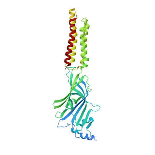
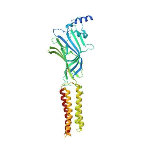
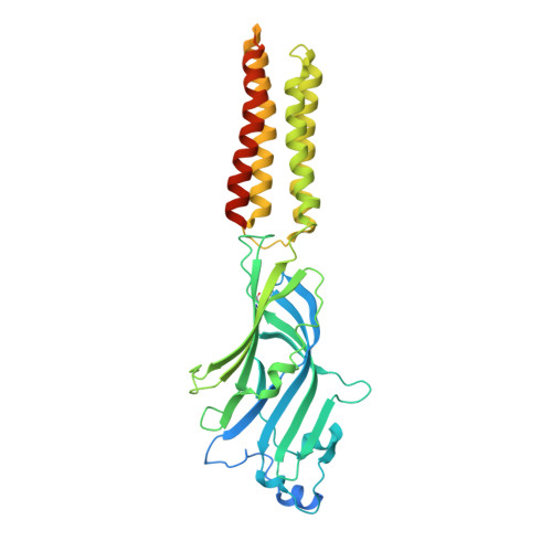
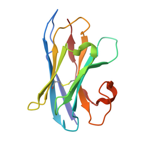
Cryo-EM structure of human full-length extrasynaptic alpha4beta3delta GABA(A)R in complex with GABA, histamine and nanobody Nb25 in a pre-open/closed state


Experimental Data Snapshot

  • Method: ELECTRON MICROSCOPY
  • Resolution: 2.90 Å
  • Aggregation State: PARTICLE 
  • Reconstruction Method: SINGLE PARTICLE 

wwPDB Validation   3D Report Full Report


This is version 1.2 of the entry. See complete history


Literature

Differential assembly diversifies GABA A receptor structures and signalling.

Sente, A.Desai, R.Naydenova, K.Malinauskas, T.Jounaidi, Y.Miehling, J.Zhou, X.Masiulis, S.Hardwick, S.W.Chirgadze, D.Y.Miller, K.W.Aricescu, A.R.

(2022) Nature 604: 190-194

  • DOI: https://doi.org/10.1038/s41586-022-04517-3
  • Primary Citation of Related Structures:  
    7QN5, 7QN6, 7QN7, 7QN8, 7QN9, 7QNA, 7QNB, 7QNC, 7QND, 7QNE

  • PubMed Abstract: 

    Type A γ-aminobutyric acid receptors (GABA A Rs) are pentameric ligand-gated chloride channels that mediate fast inhibitory signalling in neural circuits 1,2 and can be modulated by essential medicines including general anaesthetics and benzodiazepines 3 . Human GABA A R subunits are encoded by 19 paralogous genes that can, in theory, give rise to 495,235 receptor types. However, the principles that govern the formation of pentamers, the permutational landscape of receptors that may emerge from a subunit set and the effect that this has on GABAergic signalling remain largely unknown. Here we use cryogenic electron microscopy to determine the structures of extrasynaptic GABA A Rs assembled from α4, β3 and δ subunits, and their counterparts incorporating γ2 instead of δ subunits. In each case, we identified two receptor subtypes with distinct stoichiometries and arrangements, all four differing from those previously observed for synaptic, α1-containing receptors 4-7 . This, in turn, affects receptor responses to physiological and synthetic modulators by creating or eliminating ligand-binding sites at subunit interfaces. We provide structural and functional evidence that selected GABA A R arrangements can act as coincidence detectors, simultaneously responding to two neurotransmitters: GABA and histamine. Using assembly simulations and single-cell RNA sequencing data 8,9 , we calculated the upper bounds for receptor diversity in recombinant systems and in vivo. We propose that differential assembly is a pervasive mechanism for regulating the physiology and pharmacology of GABA A Rs.


  • Organizational Affiliation

    MRC Laboratory of Molecular Biology, Cambridge, UK. asente@mrc-lmb.cam.ac.uk.


Macromolecules
Find similar proteins by:  (by identity cutoff)  |  3D Structure
Entity ID: 1
MoleculeChains Sequence LengthOrganismDetailsImage
Gamma-aminobutyric acid receptor subunit alpha-4554Homo sapiensMutation(s): 0 
Gene Names: GABRA4
Membrane Entity: Yes 
UniProt & NIH Common Fund Data Resources
Find proteins for P48169 (Homo sapiens)
Explore P48169 
Go to UniProtKB:  P48169
PHAROS:  P48169
GTEx:  ENSG00000109158 
Entity Groups  
Sequence Clusters30% Identity50% Identity70% Identity90% Identity95% Identity100% Identity
UniProt GroupP48169
Glycosylation
Glycosylation Sites: 2Go to GlyGen: P48169-1
Sequence Annotations
Expand
  • Reference Sequence
Find similar proteins by:  (by identity cutoff)  |  3D Structure
Entity ID: 2
MoleculeChains Sequence LengthOrganismDetailsImage
Gamma-aminobutyric acid receptor subunit beta-3
B, C, D
473Homo sapiensMutation(s): 0 
Gene Names: GABRB3
Membrane Entity: Yes 
UniProt & NIH Common Fund Data Resources
Find proteins for P28472 (Homo sapiens)
Explore P28472 
Go to UniProtKB:  P28472
PHAROS:  P28472
GTEx:  ENSG00000166206 
Entity Groups  
Sequence Clusters30% Identity50% Identity70% Identity90% Identity95% Identity100% Identity
UniProt GroupP28472
Glycosylation
Glycosylation Sites: 2Go to GlyGen: P28472-1
Sequence Annotations
Expand
  • Reference Sequence
Find similar proteins by:  (by identity cutoff)  |  3D Structure
Entity ID: 3
MoleculeChains Sequence LengthOrganismDetailsImage
Gamma-aminobutyric acid receptor subunit delta472Homo sapiensMutation(s): 0 
Gene Names: GABRD
Membrane Entity: Yes 
UniProt & NIH Common Fund Data Resources
Find proteins for O14764 (Homo sapiens)
Explore O14764 
Go to UniProtKB:  O14764
PHAROS:  O14764
GTEx:  ENSG00000187730 
Entity Groups  
Sequence Clusters30% Identity50% Identity70% Identity90% Identity95% Identity100% Identity
UniProt GroupO14764
Glycosylation
Glycosylation Sites: 1Go to GlyGen: O14764-1
Sequence Annotations
Expand
  • Reference Sequence
Find similar proteins by:  (by identity cutoff)  |  3D Structure
Entity ID: 4
MoleculeChains Sequence LengthOrganismDetailsImage
Nanobody Nb25F [auth L],
G [auth M]
121Homo sapiensMutation(s): 0 
Entity Groups  
Sequence Clusters30% Identity50% Identity70% Identity90% Identity95% Identity100% Identity
Sequence Annotations
Expand
  • Reference Sequence
Oligosaccharides

Help

Entity ID: 5
MoleculeChains Length2D Diagram Glycosylation3D Interactions
beta-D-mannopyranose-(1-4)-2-acetamido-2-deoxy-beta-D-glucopyranose-(1-4)-2-acetamido-2-deoxy-beta-D-glucopyranoseH [auth a]3N-Glycosylation
Glycosylation Resources
GlyTouCan:  G15407YE
GlyCosmos:  G15407YE
GlyGen:  G15407YE
Entity ID: 6
MoleculeChains Length2D Diagram Glycosylation3D Interactions
2-acetamido-2-deoxy-beta-D-glucopyranose-(1-4)-2-acetamido-2-deoxy-beta-D-glucopyranoseI [auth b],
K [auth c],
M [auth d]
2N-Glycosylation
Glycosylation Resources
GlyTouCan:  G42666HT
GlyCosmos:  G42666HT
GlyGen:  G42666HT
Entity ID: 7
MoleculeChains Length2D Diagram Glycosylation3D Interactions
alpha-D-mannopyranose-(1-3)-[alpha-D-mannopyranose-(1-6)]beta-D-mannopyranose-(1-4)-2-acetamido-2-deoxy-beta-D-glucopyranose-(1-4)-2-acetamido-2-deoxy-beta-D-glucopyranoseJ [auth F],
L [auth G],
N [auth H]
5N-Glycosylation
Glycosylation Resources
GlyTouCan:  G22768VO
GlyCosmos:  G22768VO
GlyGen:  G22768VO
Small Molecules
Ligands 4 Unique
IDChains Name / Formula / InChI Key2D Diagram3D Interactions
R16
Query on R16

Download Ideal Coordinates CCD File 
Q [auth B]HEXADECANE
C16 H34
DCAYPVUWAIABOU-UHFFFAOYSA-N
NAG
Query on NAG

Download Ideal Coordinates CCD File 
O [auth A],
U [auth E]
2-acetamido-2-deoxy-beta-D-glucopyranose
C8 H15 N O6
OVRNDRQMDRJTHS-FMDGEEDCSA-N
D10
Query on D10

Download Ideal Coordinates CCD File 
S [auth D]DECANE
C10 H22
DIOQZVSQGTUSAI-UHFFFAOYSA-N
HSM (Subject of Investigation/LOI)
Query on HSM

Download Ideal Coordinates CCD File 
P [auth B],
R [auth C],
T [auth D]
HISTAMINE
C5 H9 N3
NTYJJOPFIAHURM-UHFFFAOYSA-N
Experimental Data & Validation

Experimental Data

  • Method: ELECTRON MICROSCOPY
  • Resolution: 2.90 Å
  • Aggregation State: PARTICLE 
  • Reconstruction Method: SINGLE PARTICLE 
EM Software:
TaskSoftware PackageVersion
MODEL REFINEMENTREFMAC5
MODEL REFINEMENTPHENIX1.19.2
RECONSTRUCTIONRELION3.1

Structure Validation

View Full Validation Report


Entry History & Funding Information

Deposition Data


Funding OrganizationLocationGrant Number
Medical Research Council (MRC, United Kingdom)United KingdomMR/L009609/1
Medical Research Council (MRC, United Kingdom)United KingdomMC_UP_1201/15
National Institutes of Health/National Institute of General Medical Sciences (NIH/NIGMS)United Kingdom1R01-GM135550
Wellcome TrustUnited Kingdom206171/Z/17/Z
Wellcome TrustUnited Kingdom202905/Z/16/Z

Revision History  (Full details and data files)

  • Version 1.0: 2022-04-13
    Type: Initial release
  • Version 1.1: 2022-04-20
    Changes: Database references
  • Version 1.2: 2024-11-20
    Changes: Data collection, Structure summary