7PVD | pdb_00007pvd

Structure of the membrane soluble spike complex from the Lassa virus in a C1-symmetric map focused on the ectodomain


Experimental Data Snapshot

  • Method: ELECTRON MICROSCOPY
  • Resolution: 3.70 Å
  • Aggregation State: PARTICLE 
  • Reconstruction Method: SINGLE PARTICLE 

wwPDB Validation 3D Report Full Report

Validation slider image for 7PVD

This is version 1.3 of the entry. See complete history

Re-refinement Note

This entry reflects an alternative modeling of the original data in: 7PUY

Literature

Structure and receptor recognition by the Lassa virus spike complex.

Katz, M.Weinstein, J.Eilon-Ashkenazy, M.Gehring, K.Cohen-Dvashi, H.Elad, N.Fleishman, S.J.Diskin, R.

(2022) Nature 603: 174-179

  • DOI: https://doi.org/10.1038/s41586-022-04429-2
  • Primary Citation Related Structures: 
    7PUY, 7PVD

  • PubMed Abstract: 

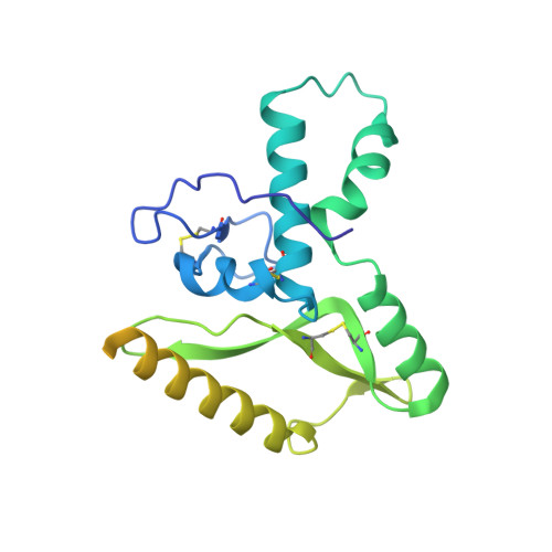
    Lassa virus (LASV) is a human pathogen, causing substantial morbidity and mortality 1,2 . Similar to other Arenaviridae, it presents a class-I spike complex on its surface that facilitates cell entry. The virus's cellular receptor is matriglycan, a linear carbohydrate that is present on α-dystroglycan 3,4 , but the molecular mechanism that LASV uses to recognize this glycan is unknown. In addition, LASV and other arenaviruses have a unique signal peptide that forms an integral and functionally important part of the mature spike 5-8 ; yet the structure, function and topology of the signal peptide in the membrane remain uncertain 9-11 . Here we solve the structure of a complete native LASV spike complex, finding that the signal peptide crosses the membrane once and that its amino terminus is located in the extracellular region. Together with a double-sided domain-switching mechanism, the signal peptide helps to stabilize the spike complex in its native conformation. This structure reveals that the LASV spike complex is preloaded with matriglycan, suggesting the mechanism of binding and rationalizing receptor recognition by α-dystroglycan-tropic arenaviruses. This discovery further informs us about the mechanism of viral egress and may facilitate the rational design of novel therapeutics that exploit this binding site.


  • Organizational Affiliation
    • Department of Chemical and Structural Biology, Weizmann Institute of Science, Rehovot, Israel.

Macromolecule Content 

  • Total Structure Weight: 179.92 kDa 
  • Atom Count: 8,902 
  • Modeled Residue Count: 1,035 
  • Deposited Residue Count: 1,509 
  • Unique protein chains: 2

Macromolecules

Find similar proteins by:|  3D Structure
Entity ID: 1
MoleculeChains  Sequence LengthOrganismDetailsImage
Glycoprotein G2A [auth a],
B [auth b],
C [auth c]
244Lassa virus JosiahMutation(s): 0 
Gene Names: GPCGP-C
UniProt
Find proteins for P08669 (Lassa virus (strain Mouse/Sierra Leone/Josiah/1976))
Explore P08669 
Go to UniProtKB:  P08669
Entity Groups
Sequence Clusters30% Identity50% Identity70% Identity90% Identity95% Identity100% Identity
UniProt GroupP08669
Glycosylation
Glycosylation Sites: 2
Sequence Annotations
Expand
Reference Sequence
Find similar proteins by:|  3D Structure
Entity ID: 2
MoleculeChains  Sequence LengthOrganismDetailsImage
Pre-glycoprotein polyprotein GP complexD [auth A],
E [auth B],
F [auth C]
259Lassa virus JosiahMutation(s): 0 
Gene Names: GPCGP-C
UniProt
Find proteins for P08669 (Lassa virus (strain Mouse/Sierra Leone/Josiah/1976))
Explore P08669 
Go to UniProtKB:  P08669
Entity Groups
Sequence Clusters30% Identity50% Identity70% Identity90% Identity95% Identity100% Identity
UniProt GroupP08669
Glycosylation
Glycosylation Sites: 5
Sequence Annotations
Expand
Reference Sequence

Oligosaccharides

Help  
Entity ID: 3
MoleculeChains Length2D Diagram GlycosylationD Interactions
2-acetamido-2-deoxy-beta-D-glucopyranose-(1-4)-2-acetamido-2-deoxy-beta-D-glucopyranose
G [auth D],
H [auth E],
I [auth F],
J [auth G],
K [auth H],
G [auth D],
H [auth E],
I [auth F],
J [auth G],
K [auth H],
L [auth I],
M [auth J],
N [auth K],
O [auth L]
2N-Glycosylation
Glycosylation Resources
GlyTouCan: G42666HT
GlyCosmos: G42666HT
GlyGen: G42666HT
Entity ID: 4
MoleculeChains Length2D Diagram GlycosylationD Interactions
alpha-D-xylopyranose-(1-3)-beta-D-glucopyranuronic acid-(1-3)-alpha-D-xylopyranose-(1-3)-beta-D-glucopyranuronic acid-(1-3)-alpha-D-xylopyranose-(1-3)-beta-D-glucopyranuronic acid-(1-3)-alpha-D-xylopyranose-(1-3)-beta-D-glucopyranuronic acid-(1-3)-alpha-D-xylopyranose-(1-3)-beta-D-glucopyranuronic acid-(1-3)-alpha-D-xylopyranose-(1-3)-beta-D-glucopyranuronic acid-(1-3)-alpha-D-xylopyranoseP [auth M]13N/A
Glycosylation Resources
GlyTouCan: G33213YN
GlyCosmos: G33213YN

Small Molecules

Ligands 1 Unique
IDChains Name / Formula / InChI Key2D Diagram3D Interactions
NAG

Query on NAG


Download:Ideal Coordinates CCD File
AA [auth C]
BA [auth C]
Q [auth a]
R [auth a]
S [auth b]
AA [auth C],
BA [auth C],
Q [auth a],
R [auth a],
S [auth b],
T [auth b],
U [auth c],
V [auth c],
W [auth A],
X [auth A],
Y [auth B],
Z [auth B]
2-acetamido-2-deoxy-beta-D-glucopyranose
C8 H15 N O6
OVRNDRQMDRJTHS-FMDGEEDCSA-N

Experimental Data & Validation

Experimental Data

  • Method: ELECTRON MICROSCOPY
  • Resolution: 3.70 Å
  • Aggregation State: PARTICLE 
  • Reconstruction Method: SINGLE PARTICLE 

Structure Validation

View Full Validation Report


Entry History 

& Funding Information

Deposition Data


Funding OrganizationLocationGrant Number
Not funded--

Revision History  (Full details and data files)

  • Version 1.0: 2021-12-29
    Type: Initial release
  • Version 1.1: 2022-03-02
    Changes: Database references
  • Version 1.2: 2022-03-16
    Changes: Database references
  • Version 1.3: 2024-11-20
    Changes: Data collection, Refinement description, Structure summary