7OJU | pdb_00007oju

Chaetomium thermophilum Naa50 GNAT-domain in complex with bisubstrate analogue CoA-Ac-MVNAL


Experimental Data Snapshot

  • Method: X-RAY DIFFRACTION
  • Resolution: 1.10 Å
  • R-Value Free: 
    0.165 (Depositor), 0.166 (DCC) 
  • R-Value Work: 
    0.145 (Depositor), 0.145 (DCC) 
  • R-Value Observed: 
    0.146 (Depositor) 

Starting Model: experimental
View more details

wwPDB Validation 3D Report Full Report

Validation slider image for 7OJU

Ligand Structure Quality Assessment 


This is version 1.3 of the entry. See complete history

Literature

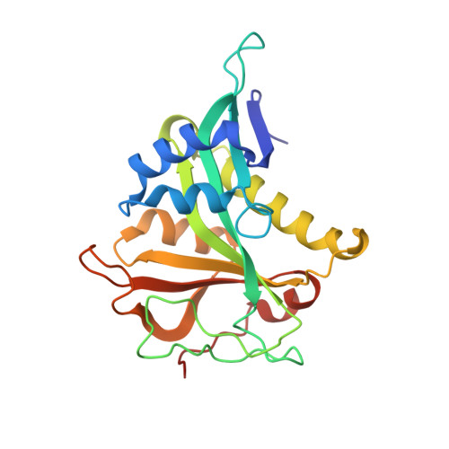
Extended N-Terminal Acetyltransferase Naa50 in Filamentous Fungi Adds to Naa50 Diversity.

Weidenhausen, J.Kopp, J.Ruger-Herreros, C.Stein, F.Haberkant, P.Lapouge, K.Sinning, I.

(2022) Int J Mol Sci 23

  • DOI: https://doi.org/10.3390/ijms231810805
  • Primary Citation Related Structures: 
    7OJU

  • PubMed Abstract: 

    Most eukaryotic proteins are N-terminally acetylated by a set of Nα acetyltransferases (NATs). This ancient and ubiquitous modification plays a fundamental role in protein homeostasis, while mutations are linked to human diseases and phenotypic defects. In particular, Naa50 features species-specific differences, as it is inactive in yeast but active in higher eukaryotes. Together with NatA, it engages in NatE complex formation for cotranslational acetylation. Here, we report Naa50 homologs from the filamentous fungi Chaetomium thermophilum and Neurospora crassa with significant N- and C-terminal extensions to the conserved GNAT domain. Structural and biochemical analyses show that Ct Naa50 shares the GNAT structure and substrate specificity with other homologs. However, in contrast to previously analyzed Naa50 proteins, it does not form NatE. The elongated N-terminus increases Naa50 thermostability and binds to dynein light chain protein 1, while our data suggest that conserved positive patches in the C-terminus allow for ribosome binding independent of NatA. Our study provides new insights into the many facets of Naa50 and highlights the diversification of NATs during evolution.


  • Organizational Affiliation
    • Heidelberg University Biochemistry Center (BZH), 69120 Heidelberg, Germany.

Macromolecule Content 

  • Total Structure Weight: 25.85 kDa 
  • Atom Count: 2,086 
  • Modeled Residue Count: 219 
  • Deposited Residue Count: 221 
  • Unique protein chains: 2

Macromolecules

Find similar proteins by:|  3D Structure
Entity ID: 1
MoleculeChains  Sequence LengthOrganismDetailsImage
N-acetyltransferase-like protein216Thermochaetoides thermophila DSM 1495Mutation(s): 0 
Gene Names: CTHT_0014970
UniProt
Find proteins for G0S1V5 (Chaetomium thermophilum (strain DSM 1495 / CBS 144.50 / IMI 039719))
Explore G0S1V5 
Go to UniProtKB:  G0S1V5
Entity Groups
Sequence Clusters30% Identity50% Identity70% Identity90% Identity95% Identity100% Identity
UniProt GroupG0S1V5
Sequence Annotations
Expand
Reference Sequence
Find similar proteins by:  Sequence   |   3D Structure  
Entity ID: 2
MoleculeChains  Sequence LengthOrganismDetailsImage
MVNAL PeptideB [auth H]5synthetic constructMutation(s): 0 

Small Molecules

Ligands 3 Unique
IDChains Name / Formula / InChI Key2D Diagram3D Interactions
CMC
(Subject of Investigation/LOI)

Query on CMC


Download:Ideal Coordinates CCD File
I [auth H]CARBOXYMETHYL COENZYME *A
C23 H38 N7 O18 P3 S
OBUOSIHPWVNVJN-GRFIIANRSA-N
P6G

Query on P6G


Download:Ideal Coordinates CCD File
H [auth A]HEXAETHYLENE GLYCOL
C12 H26 O7
IIRDTKBZINWQAW-UHFFFAOYSA-N
GOL

Query on GOL


Download:Ideal Coordinates CCD File
C [auth A],
D [auth A],
E [auth A],
F [auth A],
G [auth A]
GLYCEROL
C3 H8 O3
PEDCQBHIVMGVHV-UHFFFAOYSA-N

Experimental Data & Validation

Experimental Data

  • Method: X-RAY DIFFRACTION
  • Resolution: 1.10 Å
  • R-Value Free:  0.165 (Depositor), 0.166 (DCC) 
  • R-Value Work:  0.145 (Depositor), 0.145 (DCC) 
  • R-Value Observed: 0.146 (Depositor) 
Space Group: P 21 21 21
Unit Cell:
Length ( Å )Angle ( ˚ )
a = 36.936α = 90
b = 43.649β = 90
c = 140.065γ = 90
Software Package:
Software NamePurpose
PHENIXrefinement
XDSdata reduction
Aimlessdata scaling
PHASERphasing
PDB_EXTRACTdata extraction

Structure Validation

View Full Validation Report


Ligand Structure Quality Assessment 


Entry History 

& Funding Information

Deposition Data


Funding OrganizationLocationGrant Number
German Research Foundation (DFG)GermanySI 586/6-1
German Research Foundation (DFG)Germany201348542 - SFB 1036(TP22)

Revision History  (Full details and data files)

  • Version 1.0: 2022-09-28
    Type: Initial release
  • Version 1.1: 2022-10-05
    Changes: Database references
  • Version 1.2: 2024-01-31
    Changes: Data collection, Refinement description
  • Version 1.3: 2024-10-16
    Changes: Structure summary