7OI1 | pdb_00007oi1

Crystal structure of Synechocystis sp PCC6803 guanidinium hydrolase


Experimental Data Snapshot

  • Method: X-RAY DIFFRACTION
  • Resolution: 1.90 Å
  • R-Value Free: 
    0.208 (Depositor), 0.209 (DCC) 
  • R-Value Work: 
    0.173 (Depositor), 0.173 (DCC) 
  • R-Value Observed: 
    0.174 (Depositor) 

Starting Model: experimental
View more details

wwPDB Validation 3D Report Full Report

Validation slider image for 7OI1

This is version 1.5 of the entry. See complete history

Literature

Discovery of a Ni 2+ -dependent guanidine hydrolase in bacteria.

Funck, D.Sinn, M.Fleming, J.R.Stanoppi, M.Dietrich, J.Lopez-Igual, R.Mayans, O.Hartig, J.S.

(2022) Nature 603: 515-521

  • DOI: https://doi.org/10.1038/s41586-022-04490-x
  • Primary Citation Related Structures: 
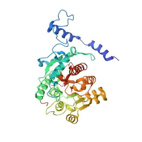
    7ESR, 7OI1

  • PubMed Abstract: 

    Nitrogen availability is a growth-limiting factor in many habitats 1 , and the global nitrogen cycle involves prokaryotes and eukaryotes competing for this precious resource. Only some bacteria and archaea can fix elementary nitrogen; all other organisms depend on the assimilation of mineral or organic nitrogen. The nitrogen-rich compound guanidine occurs widely in nature 2-4 , but its utilization is impeded by pronounced resonance stabilization 5 , and enzymes catalysing hydrolysis of free guanidine have not been identified. Here we describe the arginase family protein GdmH (Sll1077) from Synechocystis sp. PCC 6803 as a Ni 2+ -dependent guanidine hydrolase. GdmH is highly specific for free guanidine. Its activity depends on two accessory proteins that load Ni 2+ instead of the typical Mn 2+ ions into the active site. Crystal structures of GdmH show coordination of the dinuclear metal cluster in a geometry typical for arginase family enzymes and allow modelling of the bound substrate. A unique amino-terminal extension and a tryptophan residue narrow the substrate-binding pocket and identify homologous proteins in further cyanobacteria, several other bacterial taxa and heterokont algae as probable guanidine hydrolases. This broad distribution suggests notable ecological relevance of guanidine hydrolysis in aquatic habitats.


  • Organizational Affiliation
    • Department of Chemistry, University of Konstanz, Konstanz, Germany.

Macromolecule Content 

  • Total Structure Weight: 130.66 kDa 
  • Atom Count: 9,216 
  • Modeled Residue Count: 1,134 
  • Deposited Residue Count: 1,176 
  • Unique protein chains: 1

Macromolecules

Find similar proteins by:|  3D Structure
Entity ID: 1
MoleculeChains  Sequence LengthOrganismDetailsImage
Probable agmatinase 2
A, B, C
392Synechocystis sp. PCC 6803 substr. KazusaMutation(s): 0 
Gene Names: speB2sll1077
EC: 3.5.3.11 (PDB Primary Data), 3.5.3 (UniProt)
UniProt
Find proteins for P73270 (Synechocystis sp. (strain ATCC 27184 / PCC 6803 / Kazusa))
Explore P73270 
Go to UniProtKB:  P73270
Entity Groups
Sequence Clusters30% Identity50% Identity70% Identity90% Identity95% Identity100% Identity
UniProt GroupP73270
Sequence Annotations
Expand
Reference Sequence

Small Molecules

Ligands 5 Unique
IDChains Name / Formula / InChI Key2D Diagram3D Interactions
CAC

Query on CAC


Download:Ideal Coordinates CCD File
G [auth A],
N [auth B],
S [auth C]
CACODYLATE ION
C2 H6 As O2
OGGXGZAMXPVRFZ-UHFFFAOYSA-M
PEG

Query on PEG


Download:Ideal Coordinates CCD File
K [auth A]DI(HYDROXYETHYL)ETHER
C4 H10 O3
MTHSVFCYNBDYFN-UHFFFAOYSA-N
EDO

Query on EDO


Download:Ideal Coordinates CCD File
H [auth A],
I [auth A],
J [auth A],
O [auth B],
T [auth C]
1,2-ETHANEDIOL
C2 H6 O2
LYCAIKOWRPUZTN-UHFFFAOYSA-N
NI

Query on NI


Download:Ideal Coordinates CCD File
E [auth A]
F [auth A]
L [auth B]
M [auth B]
Q [auth C]
E [auth A],
F [auth A],
L [auth B],
M [auth B],
Q [auth C],
R [auth C]
NICKEL (II) ION
Ni
VEQPNABPJHWNSG-UHFFFAOYSA-N
CL

Query on CL


Download:Ideal Coordinates CCD File
D [auth A],
P [auth C]
CHLORIDE ION
Cl
VEXZGXHMUGYJMC-UHFFFAOYSA-M

Experimental Data & Validation

Experimental Data

  • Method: X-RAY DIFFRACTION
  • Resolution: 1.90 Å
  • R-Value Free:  0.208 (Depositor), 0.209 (DCC) 
  • R-Value Work:  0.173 (Depositor), 0.173 (DCC) 
  • R-Value Observed: 0.174 (Depositor) 
Space Group: C 1 2 1
Unit Cell:
Length ( Å )Angle ( ˚ )
a = 102.36α = 90
b = 140.21β = 119.75
c = 86.85γ = 90
Software Package:
Software NamePurpose
PHENIXrefinement
PHENIXrefinement
XDSdata reduction
XSCALEdata scaling
PHASERphasing

Structure Validation

View Full Validation Report


Entry History 

& Funding Information

Deposition Data


Funding OrganizationLocationGrant Number
European Research Council (ERC)Germany681777

Revision History  (Full details and data files)

  • Version 1.0: 2021-12-15
    Type: Initial release
  • Version 1.1: 2022-01-26
    Changes: Database references
  • Version 1.2: 2022-03-16
    Changes: Database references
  • Version 1.3: 2022-03-23
    Changes: Database references
  • Version 1.4: 2022-03-30
    Changes: Database references
  • Version 1.5: 2024-01-31
    Changes: Data collection, Refinement description