7NMU

Crystal structure of human platelet glycoprotein VI in complex with an inhibitory nanobody.


Experimental Data Snapshot

  • Method: X-RAY DIFFRACTION
  • Resolution: 2.50 Å
  • R-Value Free: 0.235 
  • R-Value Work: 0.174 

wwPDB Validation   3D Report Full Report


This is version 1.3 of the entry. See complete history


Literature

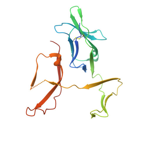
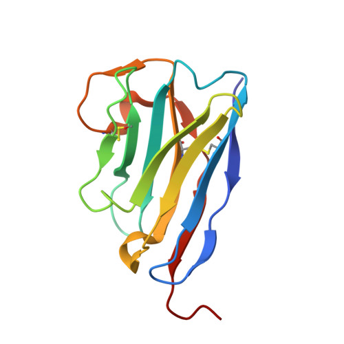
Structural characterization of a novel GPVI-nanobody complex reveals a biologically active domain-swapped GPVI dimer.

Slater, A.Di, Y.Clark, J.C.Jooss, N.J.Martin, E.M.Alenazy, F.Thomas, M.R.Ariens, R.A.S.Herr, A.B.Poulter, N.S.Emsley, J.Watson, S.P.

(2021) Blood 137: 3443-3453

  • DOI: https://doi.org/10.1182/blood.2020009440
  • Primary Citation of Related Structures:  
    7NMU

  • PubMed Abstract: 

    Glycoprotein VI (GPVI) is the major signaling receptor for collagen on platelets. We have raised 54 nanobodies (Nb), grouped into 33 structural classes based on their complementary determining region 3 loops, against recombinant GPVI-Fc (dimeric GPVI) and have characterized their ability to bind recombinant GPVI, resting and activated platelets, and to inhibit platelet activation by collagen. Nbs from 6 different binding classes showed the strongest binding to recombinant GPVI-Fc, suggesting that there was not a single dominant class. The most potent 3, Nb2, 21, and 35, inhibited collagen-induced platelet aggregation with nanomolar half maximal inhibitory concentration (IC50) values and inhibited platelet aggregation under flow. The binding KD of the most potent Nb, Nb2, against recombinant monomeric and dimeric GPVI was 0.6 and 0.7 nM, respectively. The crystal structure of monomeric GPVI in complex with Nb2 revealed a binding epitope adjacent to the collagen-related peptide (CRP) binding groove within the D1 domain. In addition, a novel conformation of GPVI involving a domain swap between the D2 domains was observed. The domain swap is facilitated by the outward extension of the C-C' loop, which forms the domain swap hinge. The functional significance of this conformation was tested by truncating the hinge region so that the domain swap cannot occur. Nb2 was still able to displace collagen and CRP binding to the mutant, but signaling was abolished in a cell-based NFAT reporter assay. This demonstrates that the C-C' loop region is important for GPVI signaling but not ligand binding and suggests the domain-swapped structure may represent an active GPVI conformation.


  • Organizational Affiliation

    Institute of Cardiovascular Sciences, College of Medical and Dental Sciences, University of Birmingham, Birmingham, United Kingdom.


Macromolecules
Find similar proteins by:  (by identity cutoff)  |  3D Structure
Entity ID: 1
MoleculeChains Sequence LengthOrganismDetailsImage
Platelet glycoprotein VIA [auth AAA],
B [auth BBB]
194Homo sapiensMutation(s): 1 
Gene Names: GP6
UniProt & NIH Common Fund Data Resources
Find proteins for Q9HCN6 (Homo sapiens)
Explore Q9HCN6 
Go to UniProtKB:  Q9HCN6
PHAROS:  Q9HCN6
GTEx:  ENSG00000088053 
Entity Groups  
Sequence Clusters30% Identity50% Identity70% Identity90% Identity95% Identity100% Identity
UniProt GroupQ9HCN6
Sequence Annotations
Expand
  • Reference Sequence
Find similar proteins by:  (by identity cutoff)  |  3D Structure
Entity ID: 2
MoleculeChains Sequence LengthOrganismDetailsImage
Nanobody 2C [auth DDD],
D [auth CCC]
130Lama glamaMutation(s): 0 
Entity Groups  
Sequence Clusters30% Identity50% Identity70% Identity90% Identity95% Identity100% Identity
Sequence Annotations
Expand
  • Reference Sequence
Small Molecules
Ligands 1 Unique
IDChains Name / Formula / InChI Key2D Diagram3D Interactions
CA
Query on CA

Download Ideal Coordinates CCD File 
E [auth CCC]CALCIUM ION
Ca
BHPQYMZQTOCNFJ-UHFFFAOYSA-N
Experimental Data & Validation

Experimental Data

  • Method: X-RAY DIFFRACTION
  • Resolution: 2.50 Å
  • R-Value Free: 0.235 
  • R-Value Work: 0.174 
  • Space Group: P 21 21 21
Unit Cell:
Length ( Å )Angle ( ˚ )
a = 69.906α = 90
b = 84.751β = 90
c = 124.041γ = 90
Software Package:
Software NamePurpose
REFMACrefinement
xia2data reduction
BUCCANEERmodel building
PHASERphasing
xia2data scaling

Structure Validation

View Full Validation Report


Entry History & Funding Information

Deposition Data


Funding OrganizationLocationGrant Number
Wellcome Trust--

Revision History  (Full details and data files)

  • Version 1.0: 2021-03-10
    Type: Initial release
  • Version 1.1: 2021-03-17
    Changes: Database references
  • Version 1.2: 2021-06-30
    Changes: Database references
  • Version 1.3: 2024-01-31
    Changes: Data collection, Database references, Derived calculations, Refinement description