Remarkable and Unexpected Mechanism for ( S )-3-Amino-4-(difluoromethylenyl)cyclohex-1-ene-1-carboxylic Acid as a Selective Inactivator of Human Ornithine Aminotransferase.
Zhu, W., Doubleday, P.F., Butrin, A., Weerawarna, P.M., Melani, R.D., Catlin, D.S., Dwight, T.A., Liu, D., Kelleher, N.L., Silverman, R.B.(2021) J Am Chem Soc 143: 8193-8207
- PubMed: 34014654 Search on PubMedSearch on PubMed Central
- DOI: https://doi.org/10.1021/jacs.1c03572
- Primary Citation Related Structures:
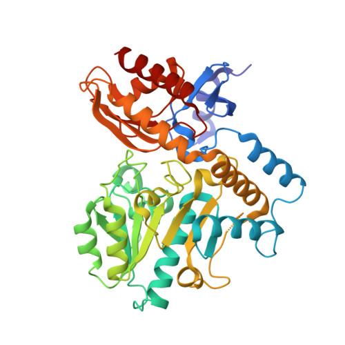
7LNM, 7LOM, 7LON - PubMed Abstract:
Human ornithine aminotransferase ( h OAT) is a pyridoxal 5'-phosphate (PLP)-dependent enzyme that was recently found to play an important role in the metabolic reprogramming of hepatocellular carcinoma (HCC) via the proline and glutamine metabolic pathways. The selective inhibition of h OAT by compound 10 exhibited potent in vivo antitumor activity. Inspired by the discovery of the aminotransferase inactivator (1 S ,3 S )-3-amino-4-(difluoromethylene)cyclopentane-1-carboxylic acid ( 5 ), we rationally designed, synthesized, and evaluated a series of six-membered-ring analogs. Among them, 14 was identified as a new selective h OAT inactivator, which demonstrated a potency 22× greater than that of 10 . Three different types of protein mass spectrometry approaches and two crystallographic approaches were employed to identify the structure of h OAT- 14 and the formation of a remarkable final adduct ( 32' ) in the active site. These spectral studies reveal an enzyme complex heretofore not observed in a PLP-dependent enzyme, which has covalent bonds to two nearby residues. Crystal soaking experiments and molecular dynamics simulations were carried out to identify the structure of the active-site intermediate 27' and elucidate the order of the two covalent bonds that formed, leading to 32' . The initial covalent reaction of the activated warhead occurs with *Thr322 from the second subunit, followed by a subsequent nucleophilic attack by the catalytic residue Lys292. The turnover mechanism of 14 by h OAT was supported by a mass spectrometric analysis of metabolites and fluoride ion release experiments. This novel mechanism for h OAT with 14 will contribute to the further rational design of selective inactivators and an understanding of potential inactivation mechanisms by aminotransferases.
- Department of Chemistry, Chemistry of Life Processes Institute, Center for Molecular Innovation and Drug Discovery, and Center for Developmental Therapeutics, Northwestern University, Evanston, Illinois 60208, United States.
Organizational Affiliation: